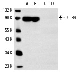
Ku-86 (B-1): sc-5280. Western blot analysis of Ku-86 expression in A-431 (A), HeLa (B), MM-142 (C) and KNRK (D) whole cell lysates. Note lack of reactivity in lanes C and D.
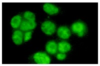
Ku-86 (B-1): sc-5280. Immunofluorescence staining of methanol-fixed HeLa cells showing nuclear staining.
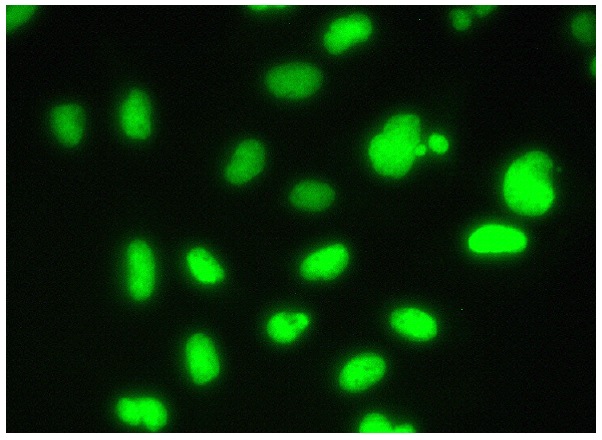
Ku-86 (B-1): sc-5280. Immunofluorescence staining of formalin-fixed HeLa cells showing nuclear localization. Kindly provided by Yang Xiang, Ph.D., Division of Newborn Medicine, Boston Childrens Hospital, Cell Biology Department, Harvard Medical School.
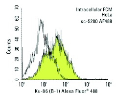
Ku-86 (B-1) Alexa Fluor 488: sc-5280 AF488. Intracellular FCM analysis of fixed and permeabilized HeLa cells. Black line histogram represents the isotype control, normal mouse IgG
1: sc-3890.
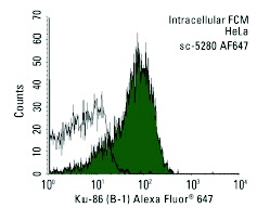
Ku-86 (B-1) Alexa Fluor 647: sc-5280 AF647. Intracellular FCM analysis of fixed and permeabilized HeLa cells. Black line histogram represents the isotype control, normal mouse IgG
1: sc-24636.
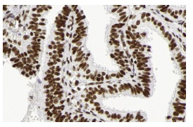
Ku-86 (B-1): sc-5280. Immunoperoxidase staining of formalin fixed, paraffin-embedded human fallopian tube tissue showing nuclear staining of glandular cells. Kindly provided by The Swedish Human Protein Atlas (HPA) program.
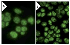
Ku-86 (B-1): sc-5280. Immunofluorescence staining of methanol-fixed HeLa cells showing nuclear localization using indirect FITC (A) staining and direct Alexa Fluor 488 (B) staining.
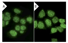
Ku-86 (B-1): sc-5280. Immunofluorescence staining of methanol-fixed HeLa cells showing nuclear localization using indirect FITC (A) staining and direct Alexa Fluor 488 (B) staining.
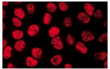
Ku-86 (B-1): sc-5280. Immunofluorescence staining of methanol-fixed HeLa cells showing nuclear localization.
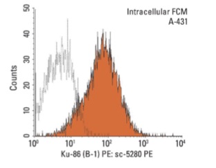
Ku-86 (B-1) PE: sc-5280 PE. Intracellular FCM analysis of fixed and permeabilized A-431 cells. Black line histogram represents the isotype control, normal mouse IgG
1: sc-2866.
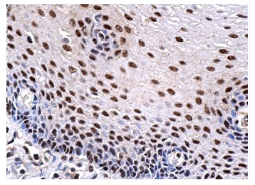
Ku-86 (B-1): sc-5280. Immunoperoxidase staining of formalin fixed, paraffin-embedded human esophagus tissue showing nuclear staining of squamous epithelial cells.


