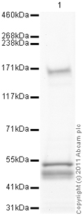
Anti-160 kD Neurofilament Medium antibody (ab64300) at 1 µg/ml + Spinal Cord (Human) Tissue Lysate - adult normal tissue (ab29188) at 10 µgSecondaryGoat Anti-Rabbit IgG H&L (HRP) preadsorbed (ab97080) at 1/5000 dilution
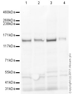
All lanes : Anti-160 kD Neurofilament Medium antibody (ab64300) at 1 µg/mlLane 1 : Spinal Cord (Mouse) Tissue LysateLane 2 : Spinal Cord (Rat) Tissue LysateLane 3 : Cerebellum Mouse Tissue LysateLane 4 : Cerebellum Rat Tissue LysateLysates/proteins at 10 µg per lane.SecondaryGoat Anti-Rabbit IgG H&L (HRP) preadsorbed (ab97080) at 1/5000 dilutionPerformed under reducing conditions.
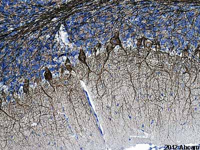
ab64300 staining rat cerebellum sections by IHC-P. The tissue was fixed with formaldehyde and a heat mediated antigen retrival step was performed with citric acid pH 6. Blocking of the sample was done with 1% BSA for 10 minutes at 21°C, followed by staining with ab64300 at 1/2000 in TBS/BSA/azide for 16h at 21°C. A biotinylated goat anti-rabbit polyclonal antibody at 1/200 was used as the secondary antibody.See Abreview
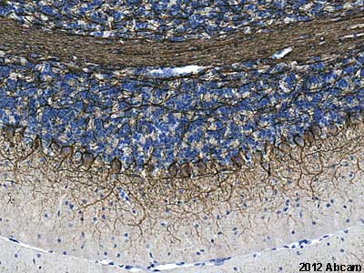
ab64300 staining mouse cerebellum sections by IHC-P. The tissue was fixed with formaldehyde and a heat mediated antigen retrival step was performed with citric acid pH 6. Blocking of the sample was done with 1% BSA for 10 minutes at 21°C, followed by staining with ab64300 at 1/2000 in TBS/BSA/azide for 16h at 21°C. A biotinylated goat anti-rabbit polyclonal antibody at 1/200 was used as the secondary antibody.See Abreview
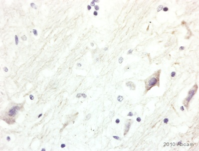
ab64300 (1/50) staining 160 kD Neurofilament Medium in paraffin-embedded Human brain tissue. Tissue underwent fixation in formaldehyde, peroxidase blocking, protein blocking and heat mediated antigen retrieval. The secondary antibody was goat anti rabbit conjugated to HRP. For further experimental details please refer to abreview. See Abreview
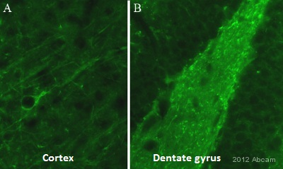
IHC-FoFr image of 160kda Neurofilament Medium staining on mouse brain sections using ab64300(1:100). The sections used came from animals perfused fixed with Paraformaldehyde 4% with 15% of a solution of saturated picric acid, in phosphate buffer 0.1M. Following postfixation in the same fixative overnight, the brains were cryoprotected in sucrose 30% overnight. Brains were then cut using a cryostat and the immunostainings were performed using the ‘free floating’ technique.See Abreview
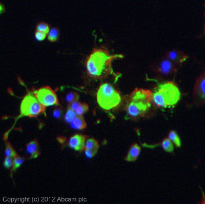
ICC/IF image of ab64300 stained PC12 cells. The cells were 4% formaldehyde fixed (10 min) and then incubated in 1%BSA / 10% normal goat serum / 0.3M glycine in 0.1% PBS-Tween for 1h to permeabilise the cells and block non-specific protein-protein interactions. The cells were then incubated with the antibody (ab64300, 5µg/ml) overnight at +4°C. The secondary antibody (green) was ab96899, DyLight® 488 goat anti-rabbit IgG (H+L) used at a 1/250 dilution for 1h. Alexa Fluor® 594 WGA was used to label plasma membranes (red) at a 1/200 dilution for 1h. DAPI was used to stain the cell nuclei (blue) at a concentration of 1.43µM.


