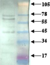
Crude cell lysate (from T-47D human breast cancer cells) was separated on 10-20% Tricine gel in denaturing conditions, then blotted and detected with 1/100 dilution of ab2508 followed by 1/5000 anti-rabbit HRP.

Crude cell lysate (from T-47D human breast cancer cells) was immunoprecipitated using ab2508 linked to anti-rabbit conjugated to magnetic beads.ab2508-precipitated proteins were separated on 10-20% Tricine gel in denaturing conditions and then blotted. The blot was treated with ab711 (1/1000) then detected using 1/5000 anti-rabbit HRP.

Composite image showing immunofuorescent staining for Laminin receptor in the rat cortex and hippocampus. Images acquired with the objective X10. Recommended dilution of antibody: 1:300-1:1000 with overnight incubation at room temperature. Free floating IHC performed with TSA amplification. Tissue was 4% perfusion PFA-fixed brain.

Immunofluorescent staining for laminin receptor in the hippocampus. The staining is observed in the cytoplasm of some astrocytes and neurons. Images acquired with X20 objective. Recommended dilution of antibody= 1:300-1:1000 with overnight incubation at room temperature. Free floating IHC performed with TSA amplification. Tissue was 4% perfusion PFA-fixed brain.

ICC/IF image of ab2508 stained human HEK 293 cells. The cells were methanol fixed (5 min), permabilised in 0.1% PBS-Tween (20 min) and incubated with the antibody (ab2508, 1:1000 dilution) for 1h at room temperature. 1%BSA / 10% normal goat serum / 0.3M glycine was used to block non-specific protein-protein interactions. The secondary antibody (green) was Alexa Fluor® 488 goat anti-rabbit IgG (H+L) used at a 1/1000 dilution for 1h. Alexa Fluor® 594 WGA was used to label plasma membranes (red). DAPI was used to stain the cell nuclei (blue). This antibody also gave a positive IF result in HeLa, HepG2 and MCF7 cells.

ICC/IF image of ab2508 stained HeLa cells. The cells were 4% PFA fixed (10 min) and then incubated in 1%BSA / 10% normal goat serum / 0.3M glycine in 0.1% PBS-Tween for 1h to permeabilise the cells and block non-specific protein-protein interactions. The cells were then incubated with the antibody (ab2508, 1/1000 dilution) overnight at +4°C. The secondary antibody (green) was Alexa Fluor® 488 goat anti-rabbit IgG (H+L) used at a 1/1000 dilution for 1h. Alexa Fluor® 594 WGA was used to label plasma membranes (red) at a 1/200 dilution for 1h. DAPI was used to stain the cell nuclei (blue) at a concentration of 1.43µM.


