Flow cytometric analysis of MCF-7 cells using AGAP8 Antibody (C-term)(green, Cat#AP20603c) compared to an isotype control of rabbit IgG(blue). AP20603c was diluted at 1:25 dilution. An Alexa Fluor® 488 goat anti-rabbit lgG at 1:400 dilution was used as the secondary antibody.

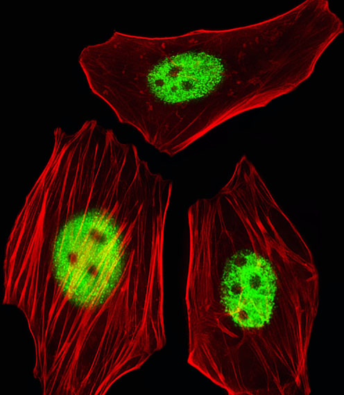
Fluorescent image of HeLa cells stained with AGAP8 Antibody (C-term)(Cat#AP20603c). AP20603c was diluted at 1:25 dilution. An Alexa Fluor 488-conjugated goat anti-rabbit lgG at 1:400 dilution was used as the secondary antibody (green). Cytoplasmic actin was counterstained with Alexa Fluor® 555 conjugated with Phalloidin (red).

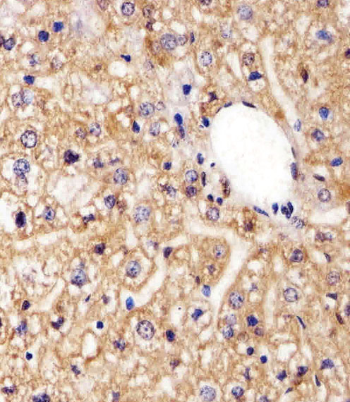
Immunohistochemical analysis of paraffin-embedded H. liver section using AGAP8 Antibody (C-term)(Cat#AP20603c). AP20603c was diluted at 1:25 dilution. A peroxidase-conjugated goat anti-rabbit IgG at 1:400 dilution was used as the secondary antibody, followed by DAB staining.

Immunohistochemical analysis of paraffin-embedded M. liver section using AGAP8 Antibody (C-term)(Cat#AP20603c). AP20603c was diluted at 1:25 dilution. A peroxidase-conjugated goat anti-rabbit IgG at 1:400 dilution was used as the secondary antibody, followed by DAB staining.

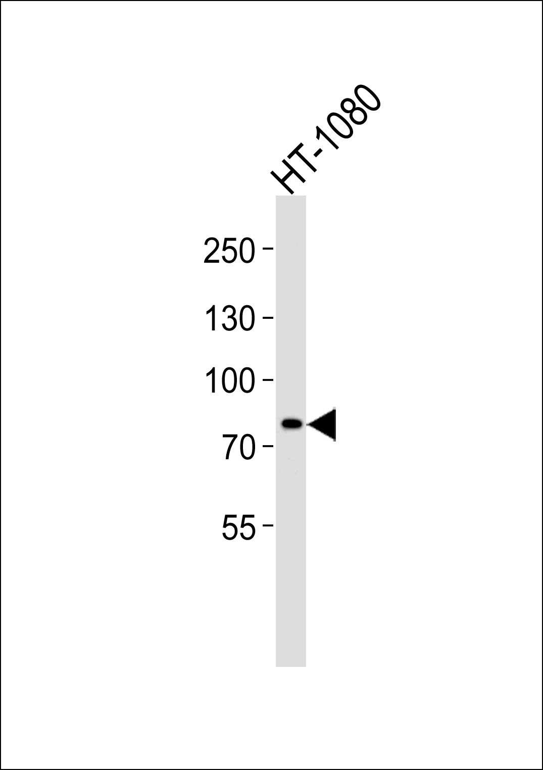
Western blot analysis of lysate from HT-1080 cell line, using AGAP8 Antibody (C-term)(Cat. # AP20603c). AP20603c was diluted at 1:1000. A goat anti-rabbit IgG H&L(HRP) at 1:5000 dilution was used as the secondary antibody. Lysate at 35ug.


