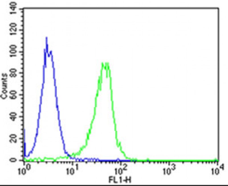
Flow cytometric analysis of Hela cells using CLN3 Antibody (Center)(green, Cat#AP20845cc) compared to an isotype control of rabbit IgG(blue). AP20845c was diluted at 1:25 dilution. An Alexa Fluor® 488 goat anti-rabbit lgG at 1:400 dilution was used as the secondary antibody.

Immunohistochemical analysis of paraffin-embedded H. liver section using CLN3 Antibody (Center)(Cat#AP20845c). AP20845c was diluted at 1:25 dilution. A undiluted biotinylated goat polyvalent antibody was used as the secondary, followed by DAB staining.

Western blot analysis of lysates from A431, KG-1, SH-SY5Y cell line, mouse kidney, mouse liver tissue (from left to right), using CLN3 Antibody (Center)(Cat. #AP20845c). AP20845c was diluted at 1:1000 at each lane. A goat anti-rabbit IgG H&L(HRP) at 1:10000 dilution was used as the secondary antibody. Lysates at 20ug per lane.