Anti-ADH5 antibody
| Name | Anti-ADH5 antibody |
|---|---|
| Supplier | Abcam |
| Catalog | ab102083 |
| Prices | $370.00 |
| Sizes | 50 µg |
| Host | Rabbit |
| Clonality | Polyclonal |
| Isotype | IgG |
| Applications | WB ICC/IF ICC/IF |
| Species Reactivities | Human, Mouse, Rat |
| Antigen | Synthetic peptide corresponding to a region within internal sequence amino acids 323-372 (KSVESVPKLV SEYMSKKIKV DEFVTHNLSF DEINKAFELM HSGKSIRTVV) of Human ADH5 (NP_000662) |
| Description | Rabbit Polyclonal |
| Gene | ADH5 |
| Conjugate | Unconjugated |
| Supplier Page | Shop |
Product images
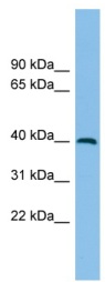 Anti-ADH5 antibody (ab102083) at 1 µg/ml + Human fetal kidney lysate at 10 µg
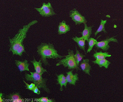 ICC/IF image of ab102083 stained HepG2 cells. The cells were 4% formaldehyde fixed (10 min) and then incubated in 1%BSA / 10% normal goat serum / 0.3M glycine in 0.1% PBS-Tween for 1h to permeabilise the cells and block non-specific protein-protein interactions. The cells were then incubated with the antibody (ab102083, 5µg/ml) overnight at +4°C. The secondary antibody (green) was ab96899, DyLight® 488 goat anti-rabbit IgG (H+L) used at a 1/250 dilution for 1h. Alexa Fluor® 594 WGA was used to label plasma membranes (red) at a 1/200 dilution for 1h. DAPI was used to stain the cell nuclei (blue) at a concentration of 1.43µM.