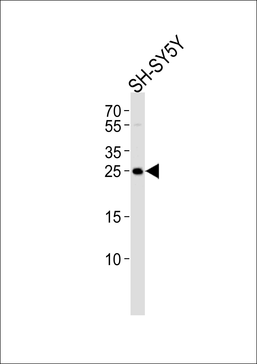
Western blot analysis of lysate from SH-SY5Y cell line, using REG3G Antibody (Center)(Cat. #AP5606c). AP5606c was diluted at 1:1000. A goat anti-rabbit IgG H&L(HRP) at 1:5000 dilution was used as the secondary antibody. Lysate at 35ug.
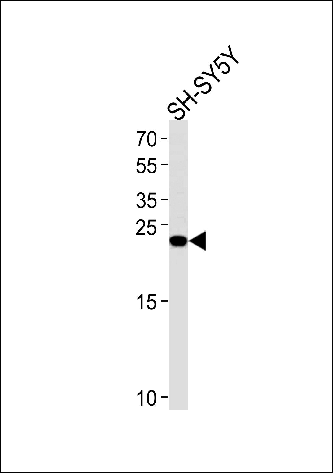
REG3G Antibody (Center) (Cat.# AP5606c) western blot analysis in SH-SY5Y cell line lysates (35ug/lane).This demonstrates the REG3G antibody detected the REG3G protein (arrow).
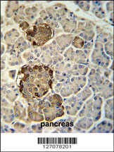
REG3G Antibody (Center) (Cat. #AP5606c) immunohistochemistry analysis in formalin fixed and paraffin embedded human pancreas tissue followed by peroxidase conjugation of the secondary antibody and DAB staining. This data demonstrates the use of the REG3G Antibody (Center) for immunohistochemistry. Clinical relevance has not been evaluated.
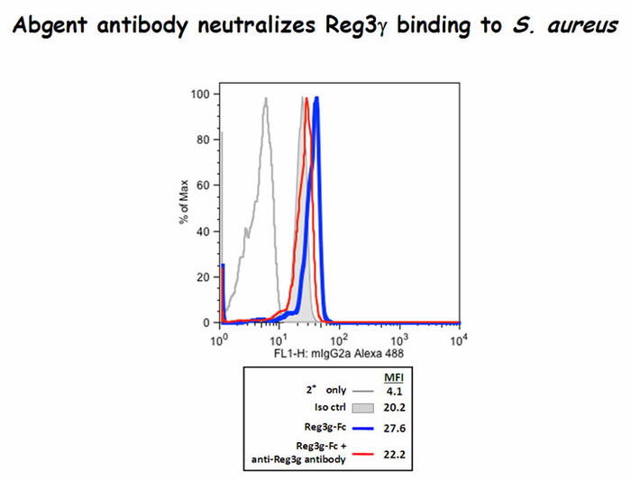
Reg3g binds to Staphylococcus aureus and the antibody did block some of this binding(Kindly offered by Dr. Choi).
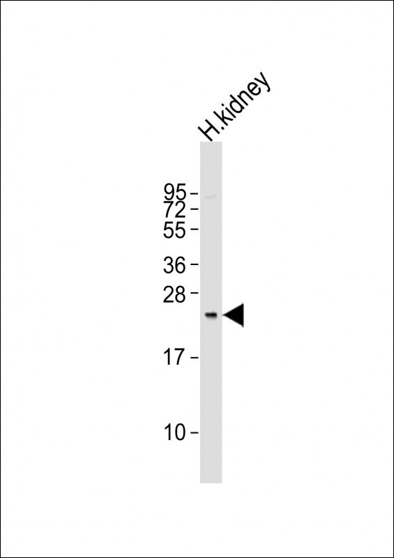
Anti-REG3G Antibody (Center)at 1:1000 dilution + human kidney lysates Lysates/proteins at 20 µg per lane. Secondary Goat Anti-Rabbit IgG, (H+L), Peroxidase conjugated at 1/10000 dilution. Predicted band size : 19 kDa Blocking/Dilution buffer: 5% NFDM/TBST.
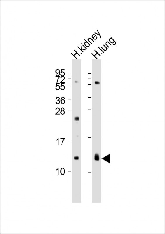
All lanes : Anti-REG3G Antibody (Center) at 1:2000 dilution Lane 1: human kidney lysates Lane 2: human lung lysates Lysates/proteins at 20 µg per lane. Secondary Goat Anti-Rabbit IgG, (H+L), Peroxidase conjugated at 1/10000 dilution. Predicted band size : 19 kDa Blocking/Dilution buffer: 5% NFDM/TBST.
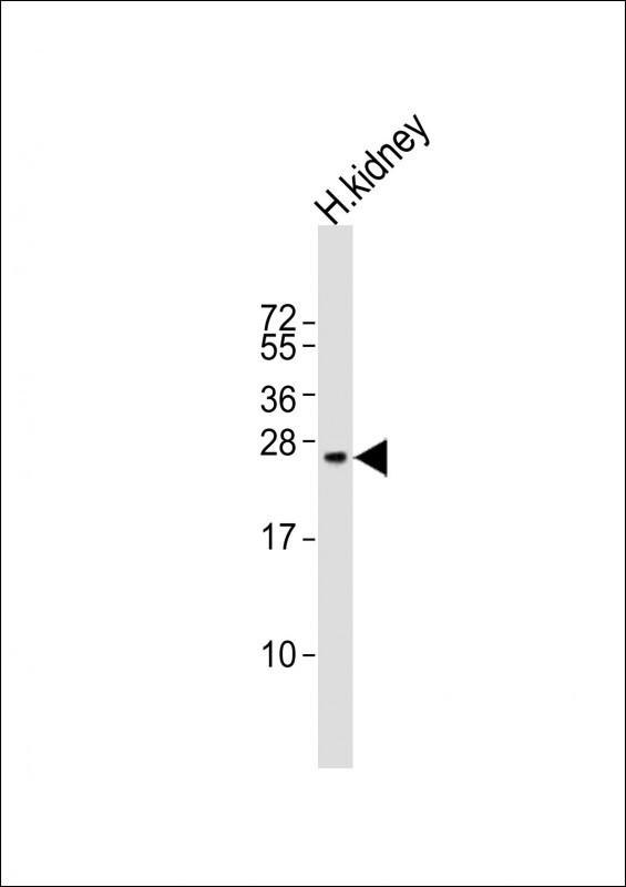
Anti-REG3G Antibody (Center)at 1:400 dilution + human kidney lysates Lysates/proteins at 20 µg per lane. Secondary Goat Anti-Rabbit IgG, (H+L), Peroxidase conjugated at 1/10000 dilution Predicted band size : 19 kDa Blocking/Dilution buffer: 5% NFDM/TBST.
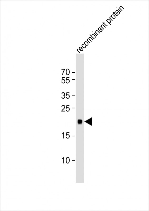
Anti-REG3G Antibody (Center)at 1:1000 dilution + recombinant protein lysates Lysates/proteins at 20 µg per lane. Secondary Goat Anti-Rabbit IgG, (H+L), Peroxidase conjugated at 1/10000 dilution Predicted band size : 19 kDa Blocking/Dilution buffer: 5% NFDM/TBST.
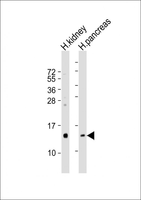
All lanes : Anti-REG3G Antibody (Center) at 1:100 dilution Lane 1: human kidney lysates Lane 2: human pancreas lysates Lysates/proteins at 20 µg per lane. Secondary Goat Anti-Rabbit IgG, (H+L), Peroxidase conjugated at 1/10000 dilution Predicted band size : 19 kDa Blocking/Dilution buffer: 5% NFDM/TBST.
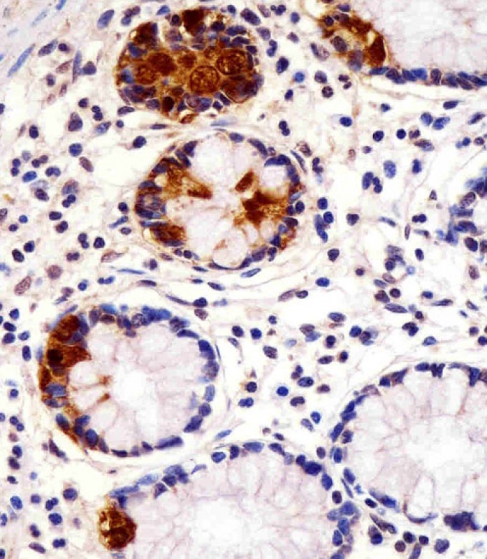
AP5606C staining REG3G in human small intestine sections by Immunohistochemistry (IHC-P - paraformaldehyde-fixed, paraffin-embedded sections). Tissue was fixed with formaldehyde and blocked with 3% BSA for 0. 5 hour at room temperature; antigen retrieval was by heat mediation with a citrate buffer (pH6). Samples were incubated with primary antibody (1/25) for 1 hours at 37°C. A undiluted biotinylated goat polyvalent antibody was used as the secondary antibody.
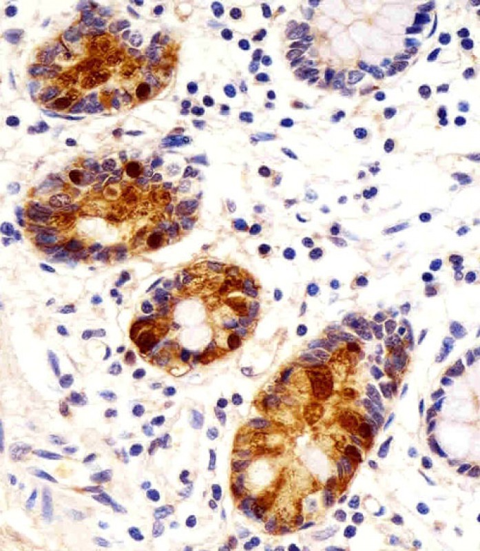
AP5606C staining REG3G in human small intestine sections by Immunohistochemistry (IHC-P - paraformaldehyde-fixed, paraffin-embedded sections). Tissue was fixed with formaldehyde and blocked with 3% BSA for 0. 5 hour at room temperature; antigen retrieval was by heat mediation with a citrate buffer (pH6). Samples were incubated with primary antibody (1/25) for 1 hours at 37°C. A undiluted biotinylated goat polyvalent antibody was used as the secondary antibody.
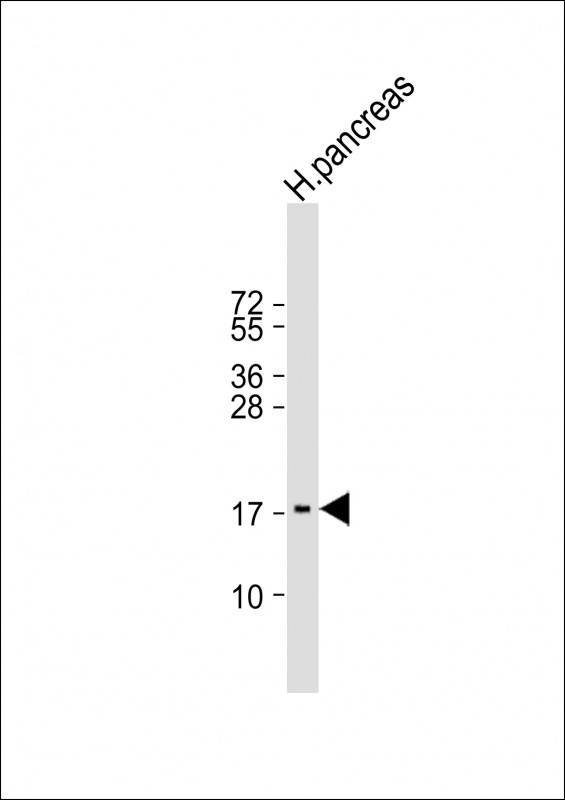
Anti-REG3G Antibody (Center) at 1:2000 dilution + human pancreas lysate Lysates/proteins at 20 µg per lane. Secondary Goat Anti-Rabbit IgG, (H+L), Peroxidase conjugated at 1/10000 dilution. Predicted band size : 19 kDa Blocking/Dilution buffer: 5% NFDM/TBST.


