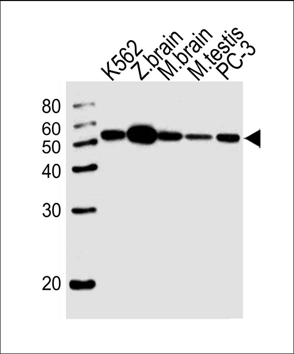
Western blot analysis of lysates from K562 cell line,zebra fish brain,mouse brain,mouse testis tissue lysate,PC-3 cell line (from left to right), using DMRTA2 Antibody (C-term)(Cat. #AW5104). AW5104 was diluted at 1:1000 at each lane. A goat anti-rabbit IgG H&L(HRP) at 1:10000 dilution was used as the secondary antibody.Lysates at 20ug per lane.

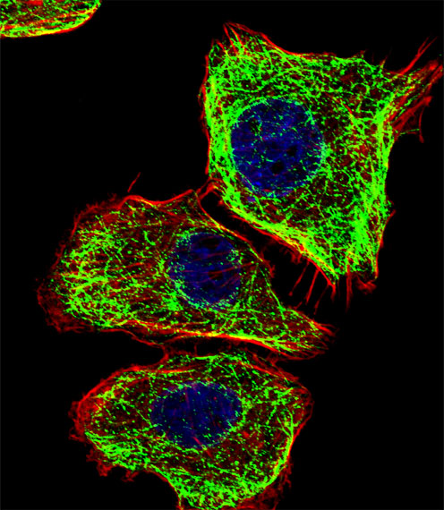
Fluorescent confocal image of U251 cell stained with DMRTA2 Antibody (C-term)(Cat#AW5104).U251 cells were fixed with 4% PFA (20 min), permeabilized with Triton X-100 (0.1%, 10 min), then incubated with DMRTA2 primary antibody (1:25, 1 h at 37℃). For secondary antibody, Alexa Fluor® 488 conjugated donkey anti-rabbit antibody (green) was used (1:400, 50 min at 37℃).Cytoplasmic actin was counterstained with Alexa Fluor® 555 (red) conjugated Phalloidin (7units/ml, 1 h at 37℃). Nuclei were counterstained with DAPI (blue) (10 µg/ml, 10 min). DMRTA2 immunoreactivity is localized to Microtubules significantly.

