COX6A2-Antibody-Center
| Name | COX6A2-Antibody-Center |
|---|---|
| Supplier | Abgent, a WuXi AppTec company |
| Catalog | AW5300 |
| Prices | $110.00, $325.00 |
| Sizes | 80 µl, 400 µl |
| Host | Rabbit |
| Clonality | Polyclonal |
| Applications | WB IHC-P |
| Species Reactivities | Human |
| Antigen | 37-66 aa |
| Purity/Format | Purified polyclonal antibody supplied in PBS with 0.09% (W/V) sodium azide. This antibody is purified through a protein A column, followed by peptide affinity purification. |
| Description | Rabbit Polyclonal |
| Gene | COX6A2 |
| Supplier Page | Shop |
Product images
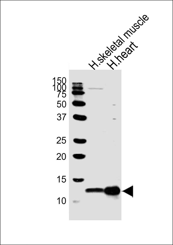 Western blot analysis of lysates from human skeletal muscle and huamn heart tissue lysate (from left to right), using COX6A2 Antibody (Center)(Cat. #AW5300). AW5300 was diluted at 1:1000 at each lane. A goat anti-rabbit IgG H&L(HRP) at 1:10000 dilution was used as the secondary antibody.
 Immunohistochemical analysis of paraffin-embedded H. skeletal muscle section using COX6A2 Antibody (Center)(Cat#AW5300). AW5300 was diluted at 1:25 dilution. A undiluted biotinylated goat polyvalent antibody was used as the secondary, followed by DAB staining.