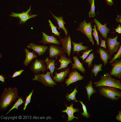
ICC/IF image of ab113503 stained HeLa cells. The cells were 100% methanol fixed (5 min) and then incubated in 1% BSA / 10% normal goat serum / 0.3M glycine in 0.1% PBS-Tween for 1h to permeabilise the cells and block non-specific protein-protein interactions. The cells were then incubated with the antibody ab113503 at 10 µg/mL overnight at +4°C. The secondary antibody (green) was DyLight® 488 Goat anti-Rabbit IgG (H+L) (ab96899) used at a 1/250 dilution for 1h. Alexa Fluor® 594 WGA was used to label plasma membranes (red) at a 1/200 dilution for 1h. DAPI was used to stain the cell nuclei (blue) at a concentration of 1.43µM.
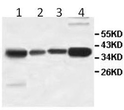
All lanes : Anti-AKR1D1 antibody (ab113503) at 1/1000 dilutionLane 1 : Human fetal testis lysateLane 2 : Human fetal colon lysateLane 3 : Human fetal intestine lysateLane 4 : HepG2 cell lysate
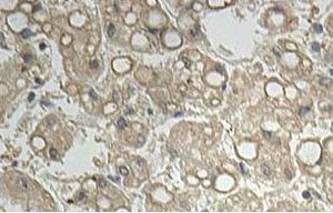
ab113503 at 1/100 dilution staining AKR1D1 in Human fetal liver by Immunohistochemistry Formalin-fixed, Paraffin-embedded tissue. Note cytoplasmic staining.