Anti-AKT1 (phospho S473) antibody [EP2109Y]
| Name | Anti-AKT1 (phospho S473) antibody [EP2109Y] |
|---|---|
| Supplier | Abcam |
| Catalog | ab81283 |
| Prices | $404.00 |
| Sizes | 100 µl |
| Host | Rabbit |
| Clonality | Monoclonal |
| Isotype | IgG |
| Clone | EP2109Y |
| Applications | IHC-F IHC-P ICC/IF WB |
| Species Reactivities | Mouse, Rat, Human |
| Antigen | A phospho specific peptide corresponding to residues surrounding Ser473 of human AKT1 |
| Blocking Peptide | AKT1 peptide |
| Description | Rabbit Monoclonal |
| Gene | AKT1 |
| Conjugate | Unconjugated |
| Supplier Page | Shop |
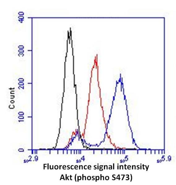
Product images
Product References
Erythroid differentiation is augmented in Reelin-deficient K562 cells and - Erythroid differentiation is augmented in Reelin-deficient K562 cells and
Chu HC, Lee HY, Huang YS, Tseng WL, Yen CJ, Cheng JC, Tseng CP. FEBS Lett. 2014 Jan 3;588(1):58-64.
Effects of tonic immobility (TI) and corticosterone (CORT) on energy status and - Effects of tonic immobility (TI) and corticosterone (CORT) on energy status and
Duan Y, Fu W, Wang S, Ni Y, Zhao R. Comp Biochem Physiol A Mol Integr Physiol. 2014 Mar;169:90-5. doi:
Targeting GRP75 improves HSP90 inhibitor efficacy by enhancing p53-mediated - Targeting GRP75 improves HSP90 inhibitor efficacy by enhancing p53-mediated
Guo W, Yan L, Yang L, Liu X, E Q, Gao P, Ye X, Liu W, Zuo J. PLoS One. 2014 Jan 17;9(1):e85766.
Frequent copy number variations of PI3K/AKT pathway and aberrant protein - Frequent copy number variations of PI3K/AKT pathway and aberrant protein
Cui W, Cai Y, Wang W, Liu Z, Wei P, Bi R, Chen W, Sun M, Zhou X. J Transl Med. 2014 Jan 13;12:10.
Low-dose benzo(a)pyrene and its epoxide metabolite inhibit myogenic - Low-dose benzo(a)pyrene and its epoxide metabolite inhibit myogenic
Chiu CY, Yen YP, Tsai KS, Yang RS, Liu SH. Toxicol Sci. 2014 Apr;138(2):344-53.
MicroRNA-21 regulates hTERT via PTEN in hypertrophic scar fibroblasts. - MicroRNA-21 regulates hTERT via PTEN in hypertrophic scar fibroblasts.
Zhu HY, Li C, Bai WD, Su LL, Liu JQ, Li Y, Shi JH, Cai WX, Bai XZ, Jia YH, Zhao B, Wu X, Li J, Hu DH. PLoS One. 2014 May 9;9(5):e97114.
NPM1 silencing reduces tumour growth and MAPK signalling in prostate cancer - NPM1 silencing reduces tumour growth and MAPK signalling in prostate cancer
Loubeau G, Boudra R, Maquaire S, Lours-Calet C, Beaudoin C, Verrelle P, Morel L. PLoS One. 2014 May 5;9(5):e96293.
Nucleolin enhances internal ribosomal entry site (IRES)-mediated translation of - Nucleolin enhances internal ribosomal entry site (IRES)-mediated translation of
Hung CY, Yang WB, Wang SA, Hsu TI, Chang WC, Hung JJ. Biochim Biophys Acta. 2014 Dec;1843(12):2843-54. doi:
The protease Omi regulates mitochondrial biogenesis through the - The protease Omi regulates mitochondrial biogenesis through the
Xu R, Hu Q, Ma Q, Liu C, Wang G. Cell Death Dis. 2014 Aug 14;5:e1373.
DNAX-activating protein 10 (DAP10) membrane adaptor associates with receptor for - DNAX-activating protein 10 (DAP10) membrane adaptor associates with receptor for
Sakaguchi M, Murata H, Aoyama Y, Hibino T, Putranto EW, Ruma IM, Inoue Y, Sakaguchi Y, Yamamoto K, Kinoshita R, Futami J, Kataoka K, Iwatsuki K, Huh NH. J Biol Chem. 2014 Aug 22;289(34):23389-402.
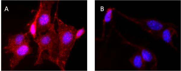
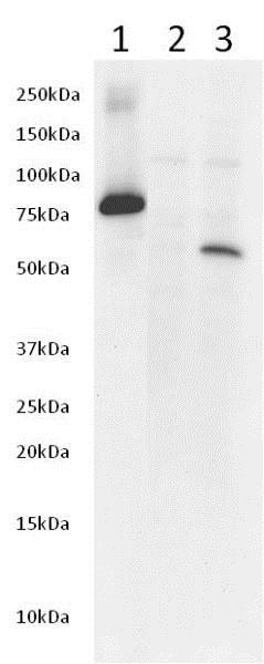
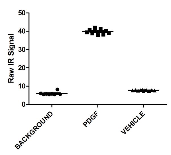
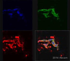
![All lanes : Anti-AKT1 (phospho S473) antibody [EP2109Y] (ab81283) at 1/10000 dilutionLane 1 : 3T3 cell lysate untreatedLane 2 : 3T3 cell lysates treated with PDGFLysates/proteins at 10 µg per lane.SecondaryHRP conjugated goat anti-rabbit at 1/2000 dilution](http://www.bioprodhub.com/system/product_images/ab_products/2/sub_1/4308_AKT1-Primary-antibodies-ab81283-2.jpg)

