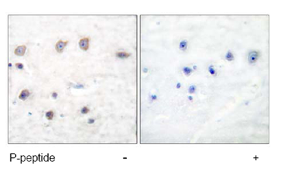
labelled with ab53093, at 1/50 dilution, staining alpha Adducin in paraffin embedded human brain tissue sections by Immunohistochemistry. Tissues had either been treated with (right image) or without (left image) the phosphopeptide.

All lanes : Anti-alpha Adducin (phospho S726) antibody (ab53093) at 1/500 dilutionLane 1 : HeLa cell extract treated with Forskolin (40nM, 30 mins) with phosphopeptideLane 2 : HeLa cell extract treated with Forskolin (40nM, 30 mins)
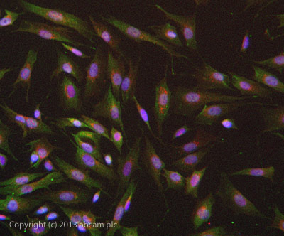
ab53093 stained SKNSH cells. The cells were 4% formaldehyde fixed (10 min) and then incubated in 1%BSA / 10% normal goat serum / 0.3M glycine in 0.1% PBS-Tween for 1h to permeabilise the cells and block non-specific protein-protein interactions. The cells were then incubated with the antibody ab53093 at 5µg/ml overnight at +4°C. The secondary antibody (green) was DyLight® 488 goat anti- rabbit (ab96899) IgG (H+L) used at a 1/1000 dilution for 1h. Alexa Fluor® 594 WGA was used to label plasma membranes (red) at a 1/200 dilution for 1h. DAPI was used to stain the cell nuclei (blue) at a concentration of 1.43µM.