Anti-alpha Adducin antibody
| Name | Anti-alpha Adducin antibody |
|---|---|
| Supplier | Abcam |
| Catalog | ab51130 |
| Prices | $387.00 |
| Sizes | 100 µg |
| Host | Rabbit |
| Clonality | Polyclonal |
| Isotype | IgG |
| Applications | IHC-P WB ELISA ICC/IF ICC/IF IP |
| Species Reactivities | Human, Mouse, Rat |
| Antigen | Synthesized non-phosphopeptide derived from human alpha Adducin around the phosphorylation site of serine 726 (T-P-SP-F-L) |
| Description | Rabbit Polyclonal |
| Gene | ADD1 |
| Conjugate | Unconjugated |
| Supplier Page | Shop |
Product images
Product References
Axon initial segment cytoskeleton comprises a multiprotein submembranous coat - Axon initial segment cytoskeleton comprises a multiprotein submembranous coat
Jones SL, Korobova F, Svitkina T. J Cell Biol. 2014 Apr 14;205(1):67-81.
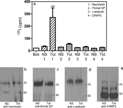
Identification of membrane-bound variant of metalloendopeptidase neurolysin (EC - Identification of membrane-bound variant of metalloendopeptidase neurolysin (EC
Wangler NJ, Santos KL, Schadock I, Hagen FK, Escher E, Bader M, Speth RC, Karamyan VT. J Biol Chem. 2012 Jan 2;287(1):114-22.


