![Anti-Apolipoprotein E antibody [EP1374Y] (HRP) (ab195855) at 1/5000 dilution + Human Plasma Total Protein Lysate at 10 µgdeveloped using the ECL techniquePerformed under reducing conditions.](http://www.bioprodhub.com/system/product_images/ab_products/2/sub_1/8539_ab195855-238103-WBab1958551.jpg)
Anti-Apolipoprotein E antibody [EP1374Y] (HRP) (ab195855) at 1/5000 dilution + Human Plasma Total Protein Lysate at 10 µgdeveloped using the ECL techniquePerformed under reducing conditions.
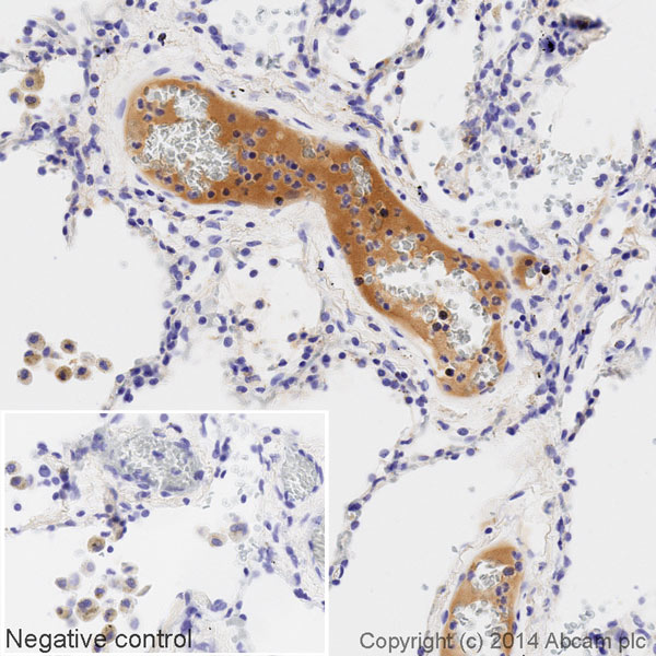
IHC image of Apolipoprotein E staining in a section of formalin-fixed paraffin-embedded normal human lung*, performed on a Leica BOND. The section was pre-treated using heat mediated antigen retrieval with sodium citrate buffer (pH6, epitope retrieval solution 1) for 20mins. The section was then incubated with ab195855, 1/50 dilution, for 15 mins at room temperature. DAB was used as the chromogen. The section was then counterstained with haematoxylin and mounted with DPX. The inset negative control image is taken from an identical assay without primary antibody.For other IHC staining systems (automated and non-automated) customers should optimize variable parameters such as antigen retrieval conditions, primary antibody concentration and antibody incubation times.*Tissue obtained from the Human Research Tissue Bank, supported by the NIHR Cambridge Biomedical Research Centre
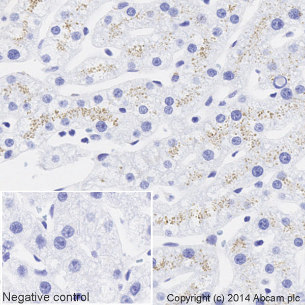
IHC image of Apolipoprotein E staining in a section of formalin-fixed paraffin-embedded normal human liver*, performed on a Leica BOND. The section was pre-treated using heat mediated antigen retrieval with sodium citrate buffer (pH6, epitope retrieval solution 1) for 20mins. The section was then incubated with ab195855, 1/71.4285714285714 dilution, for 15 mins at room temperature. DAB was used as the chromogen. The section was then counterstained with haematoxylin and mounted with DPX. The inset negative control image is taken from an identical assay without primary antibody.For other IHC staining systems (automated and non-automated) customers should optimize variable parameters such as antigen retrieval conditions, primary antibody concentration and antibody incubation times.*Tissue obtained from the Human Research Tissue Bank, supported by the NIHR Cambridge Biomedical Research Centre