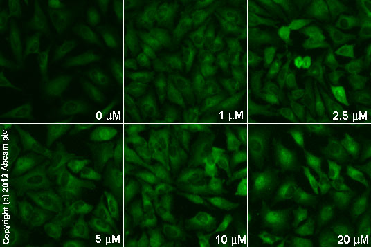
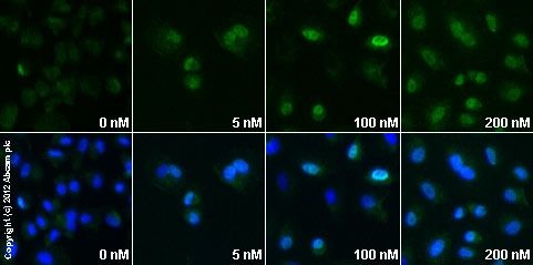
Anti-ATF3 antibody [44C3a]
| Name | Anti-ATF3 antibody [44C3a] |
|---|---|
| Supplier | Abcam |
| Catalog | ab58668 |
| Prices | $390.00 |
| Sizes | 100 µg |
| Host | Mouse |
| Clonality | Monoclonal |
| Isotype | IgG1 |
| Clone | 44C3a |
| Applications | WB DB ICC/IF ICC/IF IHC-F IHC-P |
| Species Reactivities | Rat, Human |
| Antigen | Recombinant fragment of Human ATF3 |
| Description | Mouse Monoclonal |
| Gene | ATF3 |
| Conjugate | Unconjugated |
| Supplier Page | Shop |
Product images
Product References
Satellite glial cells surrounding primary afferent neurons are activated and - Satellite glial cells surrounding primary afferent neurons are activated and
Nascimento DS, Castro-Lopes JM, Moreira Neto FL. PLoS One. 2014 Sep 23;9(9):e108152.
Differential effects of hypoxic stress in alveolar epithelial cells and - Differential effects of hypoxic stress in alveolar epithelial cells and
Signorelli S, Jennings P, Leonard MO, Pfaller W. Cell Physiol Biochem. 2010;25(1):135-44.
Inter-laboratory comparison of human renal proximal tubule (HK-2) transcriptome - Inter-laboratory comparison of human renal proximal tubule (HK-2) transcriptome
Jennings P, Aydin S, Bennett J, McBride R, Weiland C, Tuite N, Gruber LN, Perco P, Gaora PO, Ellinger-Ziegelbauer H, Ahr HJ, Kooten CV, Daha MR, Prieto P, Ryan MP, Pfaller W, McMorrow T. Toxicol In Vitro. 2009 Apr;23(3):486-99.
![Anti-ATF3 antibody [44C3a] (ab58668) + immunising recombinant protein](http://www.bioprodhub.com/system/product_images/ab_products/2/sub_1/10642_ab58668.gif)


