![Overlay histogram showing HeLa cells stained with ab89504 (red line). The cells were fixed with 80% methanol (5 min) and then permeabilized with 0.1% PBS-Tween for 20 min. The cells were then incubated in 1x PBS / 10% normal goat serum / 0.3M glycine to block non-specific protein-protein interactions followed by the antibody (ab89504, 0.1μg/1x106 cells) for 30 min at 22°C. The secondary antibody used was an anti-mouse Alexa Fluor® 488 (ab150113) at 1/2000 dilution for 30 min at 22°C. Isotype control antibody (black line) was mouse IgG1 [ICIGG1] (ab91353, 1μg/1x106 cells) used under the same conditions. Unlabelled sample (blue line) was also used as a control. Acquisition of >5,000 events were collected using a 20mW Argon ion laser (488nm) and 525/30 bandpass filter.](http://www.bioprodhub.com/system/product_images/ab_products/2/sub_1/11859_ab89504-4-ab89504FC.jpg)
Overlay histogram showing HeLa cells stained with ab89504 (red line). The cells were fixed with 80% methanol (5 min) and then permeabilized with 0.1% PBS-Tween for 20 min. The cells were then incubated in 1x PBS / 10% normal goat serum / 0.3M glycine to block non-specific protein-protein interactions followed by the antibody (ab89504, 0.1μg/1x106 cells) for 30 min at 22°C. The secondary antibody used was an anti-mouse Alexa Fluor® 488 (ab150113) at 1/2000 dilution for 30 min at 22°C. Isotype control antibody (black line) was mouse IgG1 [ICIGG1] (ab91353, 1μg/1x106 cells) used under the same conditions. Unlabelled sample (blue line) was also used as a control. Acquisition of >5,000 events were collected using a 20mW Argon ion laser (488nm) and 525/30 bandpass filter.
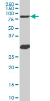
Anti-B MyB antibody (ab89504) at 5 µg/ml + K-562 cell lysate at 50 µg
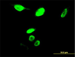
Immunofluorescence analysis of B Myb expression in PFA-fixed, permeabilized HeLa cells using ab89504 at a dilution of 10µg/ml.
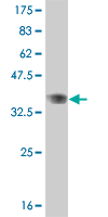
Anti-B MyB antibody (ab89504) at 5 µg/ml + immunogen (100aa protein fragment with a tag of 26kDa) at 0.2 µg