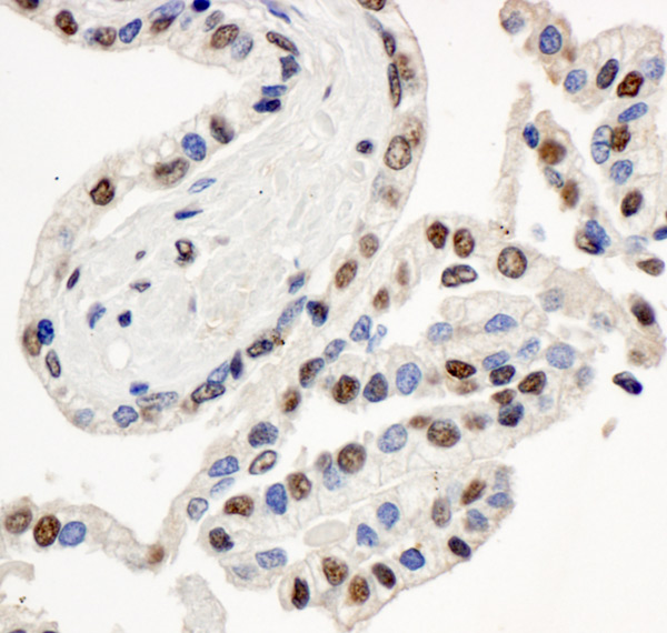
Immunohistochemistry (Formalin/PFA-fixed paraffin-embedded sections) analysis of human prostate carcinoma tissue labelling BAF57/SMARCE1 with ab70540 at 1/1000 (1µg/ml). Detection: DAB.
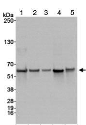
All lanes : Anti-BAF57/SMARCE1 antibody (ab70540) at 1/10000 dilutionLane 1 : Whole cell lysate from HeLa cells at 50 µgLane 2 : Whole cell lysate from HeLa cells at 15 µgLane 3 : Whole cell lysate from HeLa cells at 5 µgLane 4 : Whole cell lysate from 293T cells at 50 µgLane 5 : Whole cell lysate from NIH3T3 cells at 50 µgdeveloped using the ECL technique
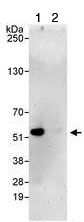
1mg of lysate from HeLa cells was immunoprecipitated using ab70540 at 3ug/mg of lysate (lane 1 or a control rabbit IgG (lane 2). For the subsequent western blot, 20% of immunoprecipitate was loaded per lane, and ab70540 was used at 1ug/ml.
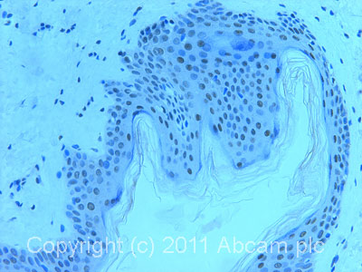
IHC image of ab70540 staining in normal human skin formalin fixed paraffin embedded tissue section, performed on a Leica BondTM system using the standard protocol F. The section was pre-treated using heat mediated antigen retrieval with sodium citrate buffer (pH6, epitope retrieval solution 1) for 20 mins. The section was then incubated with ab70540, 1µg/ml, for 15 mins at room temperature and detected using an HRP conjugated compact polymer system. DAB was used as the chromogen. The section was then counterstained with haematoxylin and mounted with DPX.For other IHC staining systems (automated and non-automated) customers should optimize variable parameters such as antigen retrieval conditions, primary antibody concentration and antibody incubation times.