![All lanes : Anti-BAF57/SMARCE1 antibody [EPR8848] (ab131328) at 1/1000 dilutionLane 1 : MCF-7 cell lysateLane 2 : HeLa cell lysateLane 3 : Jurkat cell lysateLane 4 : Raji cell lysateLysates/proteins at 10 µg per lane.SecondaryGoat anti-rabbit HRP at 1/2000 dilution](http://www.bioprodhub.com/system/product_images/ab_products/2/sub_1/12247_BAF57SMARCE1-Primary-antibodies-ab131328-1.JPG)
All lanes : Anti-BAF57/SMARCE1 antibody [EPR8848] (ab131328) at 1/1000 dilutionLane 1 : MCF-7 cell lysateLane 2 : HeLa cell lysateLane 3 : Jurkat cell lysateLane 4 : Raji cell lysateLysates/proteins at 10 µg per lane.SecondaryGoat anti-rabbit HRP at 1/2000 dilution
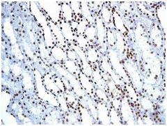
Immunohistochemical analysis of paraffin-embedded Human kidney tissue labelling BAF57/SMARCE1 with ab131328 at 1/100 dilution.
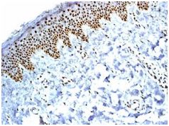
Immunohistochemical analysis of paraffin-embedded Human skin tissue labelling BAF57/SMARCE1 with ab131328 at 1/100 dilution.
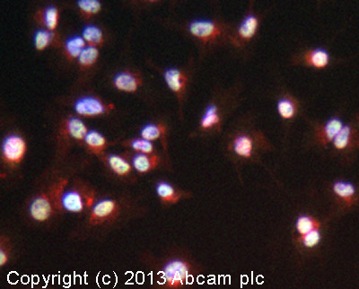
ICC/IF image of ab131328 stained HepG2 cells. The cells were 4% formaldehyde fixed (10 min) and then incubated in 1%BSA / 10% normal goat serum / 0.3M glycine in 0.1% PBS-Tween for 1h to permeabilise the cells and block non-specific protein-protein interactions. The cells were then incubated with the antibody (ab131328, 1/100 dilution) overnight at +4°C. The secondary antibody (green) was ab96899, DyLight® 488 goat anti-rabbit IgG (H+L) used at a 1/250 dilution for 1h. Alexa Fluor® 594 WGA was used to label plasma membranes (red) at a 1/200 dilution for 1h. DAPI was used to stain the cell nuclei (blue) at a concentration of 1.43µM.
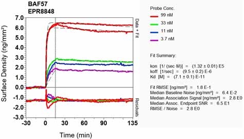
Equilibrium disassociation constant (KD)Learn more about KD Click here to learn more about KD