Anti-beta Actin antibody [mAbcam 8224] - Loading Control
| Name | Anti-beta Actin antibody [mAbcam 8224] - Loading Control |
|---|---|
| Supplier | Abcam |
| Catalog | ab8224 |
| Prices | $401.00 |
| Sizes | 100 µg |
| Host | Mouse |
| Clonality | Monoclonal |
| Isotype | IgG3 |
| Clone | mAbcam 8224 |
| Applications | FC IHC-P WB ICC/IF ICC/IF |
| Species Reactivities | Mouse, Rat, Rabbit, Chicken, Bovine, Cat, Dog, Human, Pig, Xenopus, Drosophila, S. pombe, Hamster |
| Antigen | Synthetic peptide conjugated to KLH derived from within residues 1 - 100 of Human beta Actin |
| Description | Mouse Monoclonal |
| Gene | ACTB |
| Conjugate | Unconjugated |
| Supplier Page | Shop |
Product images
Product References
Potential theranostics application of bio-synthesized silver nanoparticles - Potential theranostics application of bio-synthesized silver nanoparticles
Mukherjee S, Chowdhury D, Kotcherlakota R, Patra S, B V, Bhadra MP, Sreedhar B, Patra CR. Theranostics. 2014 Jan 29;4(3):316-35.
Silencing motifs in the Clr2 protein from fission yeast, Schizosaccharomyces - Silencing motifs in the Clr2 protein from fission yeast, Schizosaccharomyces
Steinhauf D, Rodriguez A, Vlachakis D, Virgo G, Maksimov V, Kristell C, Olsson I, Linder T, Kossida S, Bongcam-Rudloff E, Bjerling P. PLoS One. 2014 Jan 27;9(1):e86948.
Induced pluripotent stem cells as a model for telomeric abnormalities in ICF type - Induced pluripotent stem cells as a model for telomeric abnormalities in ICF type
Sagie S, Ellran E, Katzir H, Shaked R, Yehezkel S, Laevsky I, Ghanayim A, Geiger D, Tzukerman M, Selig S. Hum Mol Genet. 2014 Jul 15;23(14):3629-40.
Cleavage factor I links transcription termination to DNA damage response and - Cleavage factor I links transcription termination to DNA damage response and
Gaillard H, Aguilera A. PLoS Genet. 2014 Mar 6;10(3):e1004203.
The role of HuR in the post-transcriptional regulation of interleukin-3 in T - The role of HuR in the post-transcriptional regulation of interleukin-3 in T
Gonzalez-Feliciano JA, Hernandez-Perez M, Estrella LA, Colon-Lopez DD, Lopez A, Martinez M, Mauras-Rivera KR, Lasalde C, Martinez D, Araujo-Perez F, Gonzalez CI. PLoS One. 2014 Mar 21;9(3):e92457.
SIRT1 mediates FOXA2 breakdown by deacetylation in a nutrient-dependent manner. - SIRT1 mediates FOXA2 breakdown by deacetylation in a nutrient-dependent manner.
van Gent R, Di Sanza C, van den Broek NJ, Fleskens V, Veenstra A, Stout GJ, Brenkman AB. PLoS One. 2014 May 29;9(5):e98438.
Phosphorodiamidate morpholino oligomers suppress mutant huntingtin expression and - Phosphorodiamidate morpholino oligomers suppress mutant huntingtin expression and
Sun X, Marque LO, Cordner Z, Pruitt JL, Bhat M, Li PP, Kannan G, Ladenheim EE, Moran TH, Margolis RL, Rudnicki DD. Hum Mol Genet. 2014 Dec 1;23(23):6302-17.
Inducible, tightly regulated and growth condition-independent transcription - Inducible, tightly regulated and growth condition-independent transcription
Ottoz DS, Rudolf F, Stelling J. Nucleic Acids Res. 2014;42(17):e130.
Phosphorylation of the transcription activator CLOCK regulates progression - Phosphorylation of the transcription activator CLOCK regulates progression
Mahesh G, Jeong E, Ng FS, Liu Y, Gunawardhana K, Houl JH, Yildirim E, Amunugama R, Jones R, Allen DL, Edery I, Kim EY, Hardin PE. J Biol Chem. 2014 Jul 11;289(28):19681-93.
The yeast Cyc8-Tup1 complex cooperates with Hda1p and Rpd3p histone deacetylases - The yeast Cyc8-Tup1 complex cooperates with Hda1p and Rpd3p histone deacetylases
Fleming AB, Beggs S, Church M, Tsukihashi Y, Pennings S. Biochim Biophys Acta. 2014 Nov;1839(11):1242-55. doi:

![All lanes : Anti-beta Actin antibody [mAbcam 8224] - Loading Control (ab8224) at 1 µg/mlLane 1 : A431 (Human epithelial carcinoma cell line) Whole Cell LysateLane 2 : HEK293 (Human embryonic kidney cell line) Whole Cell LysateLane 3 : NIH 3T3 (Mouse embryonic fibroblast cell line) Whole Cell LysateLane 4 : PC12 (Rat adrenal pheochromocytoma cell line) Whole Cell LysateLane 5 : Skeletal Muscle (Human) Tissue Lysate - adult normal tissueLysates/proteins at 20 µg per lane.SecondaryGoat Anti-Mouse IgG H&L (Alexa Fluor® 790) (ab175783) at 1/10000 dilution](http://www.bioprodhub.com/system/product_images/ab_products/2/sub_1/13670_ab8224-219023-FWBab8224Retest.jpg)
![All lanes : Anti-beta Actin antibody [mAbcam 8224] - Loading Control (ab8224) at 1 µg/mlLane 1 : HeLa (Human epithelial carcinoma cell line) Whole Cell LysateLane 2 : NIH 3T3 (Mouse embryonic fibroblast cell line) Whole Cell LysateLane 3 : PC12 (Rat adrenal pheochromocytoma cell line) Whole Cell LysateLysates/proteins at 10 µg per lane.SecondaryGoat Anti-Mouse IgG H&L (HRP) preadsorbed (ab97040) at 1/50000 dilutionPerformed under reducing conditions.](http://www.bioprodhub.com/system/product_images/ab_products/2/sub_1/13671_ab8224-219467-WBAP16522682.jpg)
![All lanes : Anti-beta Actin antibody [mAbcam 8224] - Loading Control (ab8224) at 1 µg/mlLane 1 : Drosophila lysateLane 2 : S. pombe lysateLane 3 : S. cerevisiae lysate (Actin 1 - please see note)Lysates/proteins at 20 µg per lane.SecondaryRabbit Anti-Mouse IgG H&L (HRP) (ab6728) at 1/5000 dilutiondeveloped using the ECL techniquePerformed under reducing conditions.](http://www.bioprodhub.com/system/product_images/ab_products/2/sub_1/13672_ab8224_2.jpg)
![Anti-beta Actin antibody [mAbcam 8224] - Loading Control (ab8224) + Xenopus embryo lysate at 20 µgSecondaryRabbit Anti-Mouse IgG H&L (HRP) (ab6728)developed using the ECL techniquePerformed under reducing conditions.](http://www.bioprodhub.com/system/product_images/ab_products/2/sub_1/13673_ab8224_4.jpg)
![All lanes : Anti-beta Actin antibody [mAbcam 8224] - Loading Control (ab8224) at 1/1000 dilutionLane 1 : Fruit fly (Drosophila melanogastor) whole cell lysate - Female Lane 2 : Fruit fly (Drosophila melanogastor) whole cell lysate - Male Lysates/proteins at 100 µg per lane.SecondaryAn HRP-conjugated Sheep polyclonal to mouse IgG at 1/10000 dilutiondeveloped using the ECL techniquePerformed under reducing conditions.](http://www.bioprodhub.com/system/product_images/ab_products/2/sub_1/13674_beta-Actin-Primary-antibodies-ab8224-11.jpg)
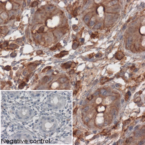
![Immunohistochemistical detection of beta Actin using antibody [mAbcam 8224] - Loading Control on formaldehyde-fixed paraffin-embedded rat cerebellum sections. Antigen retrieval step: heat mediated Citric acid pH6 buffer. Permeabilization: No. Blocking step: 1% BSA for 10 mins @ rt°C. Primary antibody dilution 1/1000 for 2 hours in TBS/BSA/azide. Secondary Antibody: anti Mouse Igs conjugated to biotin (1/200). beta Actin appears to be particularly enriched not only in the glomeruli of the Granule cell layer (indicated by red arrowheads ) but also in Microglia (indicated by green arrowheads); All positive microglia appear to be ramified thus not presumed to be activated.See Abreview](http://www.bioprodhub.com/system/product_images/ab_products/2/sub_1/13675_beta-Actin-Primary-antibodies-ab8224-13.jpg)

![Overlay histogram showing HeLa cells stained with ab8224 (red line). The cells were fixed with 80% methanol (5 min) and then permeabilized with 0.1% PBS-Tween for 20 min. The cells were then incubated in 1x PBS / 10% normal goat serum / 0.3M glycine to block non-specific protein-protein interactions followed by the antibody (ab8224, 1µg/1x106 cells) for 30 min at 22ºC. The secondary antibody used was a goat anti-mouse DyLight® 488 (IgG; H+L) (ab96879) at 1/500 dilution for 30 min at 22ºC. Isotype control antibody (black line) was Mouse IgG3 [MG3-35] (ab18394, 1µg/1x106 cells) used under the same conditions. Acquisition of >5,000 events was performed.](http://www.bioprodhub.com/system/product_images/ab_products/2/sub_1/13677_beta-Actin-Primary-antibodies-ab8224-39.jpg)
