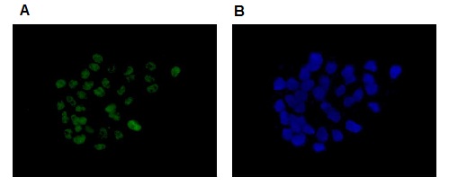
Immunocytochemistry/Immunofluorescence analysis of HepG2 (Human liver cell line) cells labelling Brd4 with purified ab128874 at 1/100 (A). Cells were fixed with 4% paraformaldehyde and permeabilized with 0.1% Triton X-100. An Alexa Fluor® 488-conjugated goat anti-rabbit IgG (1/200) was used as the secondary antibody. DAPI (blue) was used as the nuclear counter stain(B).
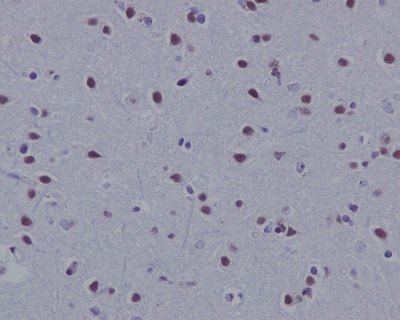
Immunohistochemistry (Formalin/PFA-fixed paraffin-embedded sections) analysis of Human brain tissue labelling Brd4 with purified ab128874 at 1/200. Heat mediated antigen retrieval was performed using Tris/EDTA buffer pH 9. An undiluted Goat Anti-Rabbit IgG H&L (HRP) was used as the secondary antibody at. Counter stained with Hematoxylin.
![Anti-Brd4 antibody [EPR5150(2)] (ab128874) at 1/1000 dilution (Purified) + NIH/3T3 Cell Lysate at 10 µgSecondaryGoat Anti-Rabbit IgG, (H+L), Peroxidase conjugated at 1/1000 dilution](http://www.bioprodhub.com/system/product_images/ab_products/2/sub_1/15501_ab128874-238986-128874-WB2.jpg)
Anti-Brd4 antibody [EPR5150(2)] (ab128874) at 1/1000 dilution (Purified) + NIH/3T3 Cell Lysate at 10 µgSecondaryGoat Anti-Rabbit IgG, (H+L), Peroxidase conjugated at 1/1000 dilution
![Anti-Brd4 antibody [EPR5150(2)] (ab128874) at 1/1000 dilution (purified) + HeLa at 10 µgSecondaryGoat Anti-Rabbit IgG, (H+L), Peroxidase conjugated at 1/1000 dilution](http://www.bioprodhub.com/system/product_images/ab_products/2/sub_1/15502_ab128874-238868-ab128874.jpg)
Anti-Brd4 antibody [EPR5150(2)] (ab128874) at 1/1000 dilution (purified) + HeLa at 10 µgSecondaryGoat Anti-Rabbit IgG, (H+L), Peroxidase conjugated at 1/1000 dilution
![Anti-Brd4 antibody [EPR5150(2)] (ab128874) at 1/200 dilution (unpurified) + HeLa at 10 µg/mlSecondaryGoat Anti-Rabbit IgG, (H+L), Peroxidase conjugated at 1/1000 dilution](http://www.bioprodhub.com/system/product_images/ab_products/2/sub_1/15503_ab128874-238867-ab128874.jpg)
Anti-Brd4 antibody [EPR5150(2)] (ab128874) at 1/200 dilution (unpurified) + HeLa at 10 µg/mlSecondaryGoat Anti-Rabbit IgG, (H+L), Peroxidase conjugated at 1/1000 dilution
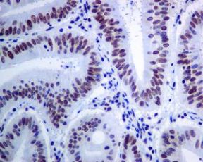
Unpurified ab128874 at 1/100 dilution staining Brd4 in Human colon carcinoma tissue by Immunohistochemistry.
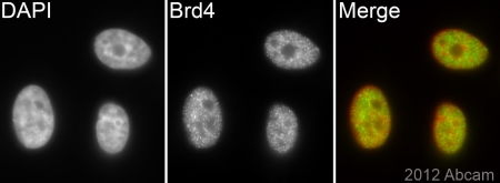
Unpurified ab128874 (1/500) staining Brd4 in Hela cells (green). Cells were fixed in paraformaldehyde, permeabilised in 0.5% Triton X100/PBS and counter stained with DAPI in order to highlight the nucleus (red). For further experimental details please refer to Abreview.See Abreview
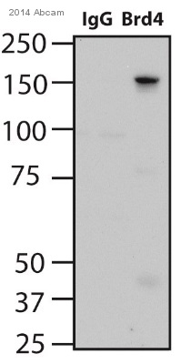
ab128874 Immunoprecipitating Brd4 in human HEK293 whole cell lysate. 1000µg of cell lysate was incubated with unpurified primary antibody (1 µg/ml in 50 mM TRIS) and matrix (Protein G) for 16 hours at 4°C. For western blotting ab131366 (anti-mouse HRP, 1/10000) was used to confirm successful immunoprecipation.See Abreview
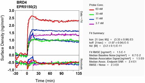
Equilibrium disassociation constant (KD)Learn more about KD Click here to learn more about KD


![Anti-Brd4 antibody [EPR5150(2)] (ab128874) at 1/1000 dilution (Purified) + NIH/3T3 Cell Lysate at 10 µgSecondaryGoat Anti-Rabbit IgG, (H+L), Peroxidase conjugated at 1/1000 dilution](http://www.bioprodhub.com/system/product_images/ab_products/2/sub_1/15501_ab128874-238986-128874-WB2.jpg)
![Anti-Brd4 antibody [EPR5150(2)] (ab128874) at 1/1000 dilution (purified) + HeLa at 10 µgSecondaryGoat Anti-Rabbit IgG, (H+L), Peroxidase conjugated at 1/1000 dilution](http://www.bioprodhub.com/system/product_images/ab_products/2/sub_1/15502_ab128874-238868-ab128874.jpg)
![Anti-Brd4 antibody [EPR5150(2)] (ab128874) at 1/200 dilution (unpurified) + HeLa at 10 µg/mlSecondaryGoat Anti-Rabbit IgG, (H+L), Peroxidase conjugated at 1/1000 dilution](http://www.bioprodhub.com/system/product_images/ab_products/2/sub_1/15503_ab128874-238867-ab128874.jpg)


