Anti-CACNG6 antibody
| Name | Anti-CACNG6 antibody |
|---|---|
| Supplier | Abcam |
| Catalog | ab170786 |
| Prices | $370.00 |
| Sizes | 400 µl |
| Host | Rabbit |
| Clonality | Polyclonal |
| Isotype | IgG |
| Applications | IHC-P WB ICC/IF ICC/IF |
| Species Reactivities | Human |
| Antigen | Synthetic peptide within Human CACNG6 aa 115-144 (internal sequence) conjugated to Keyhole Limpet Haemocyanin (KLH) |
| Description | Rabbit Polyclonal |
| Gene | CACNG6 |
| Conjugate | Unconjugated |
| Supplier Page | Shop |
Product images
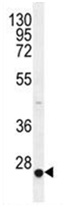 Anti-CACNG6 antibody (ab170786) at 1/100 dilution + HepG2 cell lysate at 35 µg
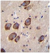 Immunohistochemical analysis of formalin fixed, paraffin embedded Human brain tissue labeling CACNG6 with ab170786 at 1/10 dilution, followed by peroxidase conjugation of the secondary antibody and DAB staining.
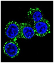 Confocal immunofluorescent analysis of HepG2 cells labeling CACNG6 with ab170786 at 1/10 dilution, followed by Alexa Fluor 488-conjugated goat anti-rabbit lgG (green). DAPI was used to stain the cell nuclear (blue).