Anti-Calcineurin A antibody
| Name | Anti-Calcineurin A antibody |
|---|---|
| Supplier | Abcam |
| Catalog | ab3673 |
| Prices | $398.00 |
| Sizes | 100 µl |
| Host | Rabbit |
| Clonality | Polyclonal |
| Isotype | IgG |
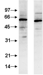
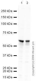
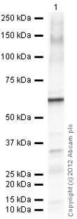
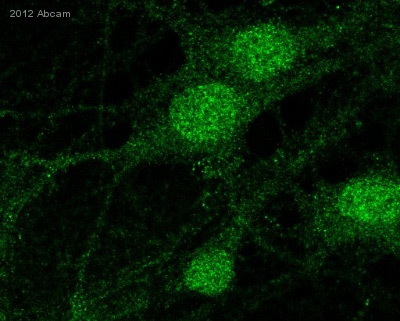
| Applications | IP WB ICC/IF ICC/IF |
| Species Reactivities | Mouse, Rat, Human |
| Antigen | Full length Calcineurin A fusion expressed in E |
| Description | Rabbit Polyclonal |
| Gene | PPP3CA |
| Conjugate | Unconjugated |
| Supplier Page | Shop |
Product images
Product References
Interaction between maternal and postnatal high fat diet leads to a greater risk - Interaction between maternal and postnatal high fat diet leads to a greater risk
Turdi S, Ge W, Hu N, Bradley KM, Wang X, Ren J. J Mol Cell Cardiol. 2013 Feb;55:117-29.
Hypertrophic cardiomyopathy in high-fat diet-induced obesity: role of suppression - Hypertrophic cardiomyopathy in high-fat diet-induced obesity: role of suppression
Fang CX, Dong F, Thomas DP, Ma H, He L, Ren J. Am J Physiol Heart Circ Physiol. 2008 Sep;295(3):H1206-H1215. doi:
The Down syndrome critical region protein RCAN1 regulates long-term potentiation - The Down syndrome critical region protein RCAN1 regulates long-term potentiation
Hoeffer CA, Dey A, Sachan N, Wong H, Patterson RJ, Shelton JM, Richardson JA, Klann E, Rothermel BA. J Neurosci. 2007 Nov 28;27(48):13161-72.


