![All lanes : Anti-Calmegin antibody [EPR11832] (ab171971) at 1/10000 dilutionLane 1 : Jurkat cell lysateLane 2 : Human testis lysateLane 3 : Mouse testis lysateLane 4 : Rat testis lysateLysates/proteins at 10 µg per lane.SecondaryStandard HRP labeled goat anti-rabbit at 1/2000 dilutiondeveloped using the ECL technique](http://www.bioprodhub.com/system/product_images/ab_products/2/sub_1/19463_ab171971-172710-ab171971wb.jpg)
All lanes : Anti-Calmegin antibody [EPR11832] (ab171971) at 1/10000 dilutionLane 1 : Jurkat cell lysateLane 2 : Human testis lysateLane 3 : Mouse testis lysateLane 4 : Rat testis lysateLysates/proteins at 10 µg per lane.SecondaryStandard HRP labeled goat anti-rabbit at 1/2000 dilutiondeveloped using the ECL technique
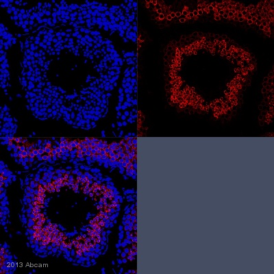
IHC-P image of Calmegin staining with ab171971 on tissue sections from adult marmoset testis. The sections were subjected to heat-mediated antigen retrieval using Dako antigen retrieval solution. The sections were then blocked with 5% milk for 30 minutes at 25°C, before incubation with ab171971 (1/500 dilution) for 18 hours at 4°C. The secondary was an Alexa-Fluor 555 conjugated goat anti-rabbit polyclonal, used at a 1/500 dilution.See Abreview
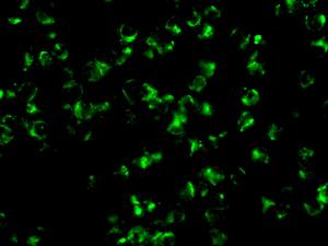
Immunofluorescence analysis of Jurkat cells, labeling Calmegin using ab171971 at a 1/50 dilution.
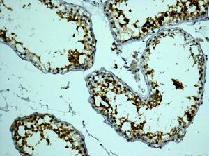
Immunohistochemical analysis of paraffin-embedded Human testis tissue, labeling Calmegin using ab171971 at a 1/100 dilution.
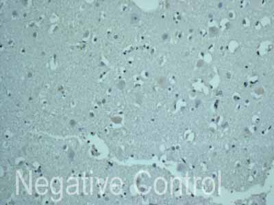
ab171971 showing -ve staining in Human normal brain tissue.
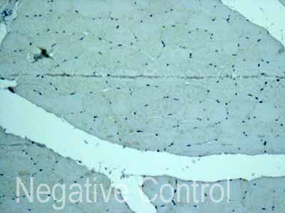
ab171971 showing -ve staining in Human skeletal muscle tissue.
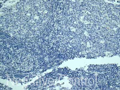
ab171971 showing -ve staining in Human normal tonsil tissue.
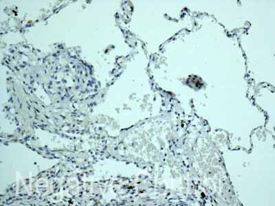
ab171971 showing -ve staining in Human normal lung tissue.
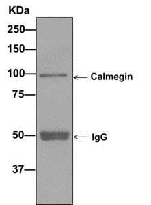
Western blot analysis on immunoprecipitation pellet from Human testis lysate, labeling Calmegin using ab171971 at a 1/10 dilution and an IgG control.