![All lanes : Anti-Calreticulin antibody [EPR3924] - ER Marker (HRP) (ab195511) at 1/5000 dilutionLane 1 : HepG2 (Human hepatocellular liver carcinoma cell line) Whole Cell LysateLane 2 : HeLa (Human epithelial carcinoma cell line) Whole Cell Lysate (ab150035)Lane 3 : Brain (Human) Tissue Lysate - fetal normal tissueLysates/proteins at 10 µg per lane.developed using the ECL techniquePerformed under reducing conditions.](http://www.bioprodhub.com/system/product_images/ab_products/2/sub_1/19847_ab195511-238041-WBab1955111.jpg)
All lanes : Anti-Calreticulin antibody [EPR3924] - ER Marker (HRP) (ab195511) at 1/5000 dilutionLane 1 : HepG2 (Human hepatocellular liver carcinoma cell line) Whole Cell LysateLane 2 : HeLa (Human epithelial carcinoma cell line) Whole Cell Lysate (ab150035)Lane 3 : Brain (Human) Tissue Lysate - fetal normal tissueLysates/proteins at 10 µg per lane.developed using the ECL techniquePerformed under reducing conditions.
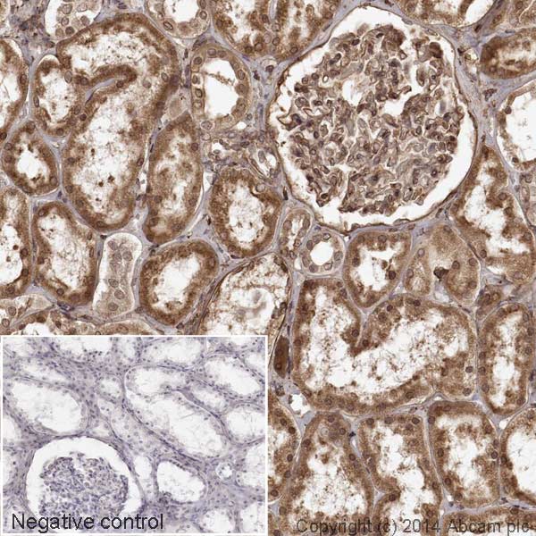
IHC image of Calreticulin staining in a section of formalin-fixed paraffin-embedded human normal kidney*. The section was pre-treated using pressure cooker heat mediated antigen retrieval with sodium citrate buffer (pH6) for 30mins, and incubated overnight at +4°C with ab195511 at 1µg/ml. DAB was used as the chromogen (ab103723), diluted 1/100 and incubated for 10min at room temperature. The section was counterstained with haematoxylin and mounted with DPX. The inset negative control image is taken from an identical assay without primary antibody.For other IHC staining systems (automated and non-automated) customers should optimize variable parameters such as antigen retrieval conditions, primary antibody concentration and antibody incubation times.*Tissue obtained from the Human Research Tissue Bank, supported by the NIHR Cambridge Biomedical Research Centre