![All lanes : Anti-CAP1 antibody [EPR8339(B)] (ab155079) at 1/1000 dilutionLane 1 : HeLa cell lysateLane 2 : 293T cell lysateLane 3 : A431 cell lysateLane 4 : HepG2 cell lysateLane 5 : Caco-2 cell lysateLysates/proteins at 10 µg per lane.](http://www.bioprodhub.com/system/product_images/ab_products/2/sub_1/20301_CAP1-Primary-antibodies-ab155079-1.jpg)
All lanes : Anti-CAP1 antibody [EPR8339(B)] (ab155079) at 1/1000 dilutionLane 1 : HeLa cell lysateLane 2 : 293T cell lysateLane 3 : A431 cell lysateLane 4 : HepG2 cell lysateLane 5 : Caco-2 cell lysateLysates/proteins at 10 µg per lane.
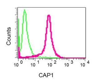
Flow cytometric analysis of permeabilized 293 cells, labeling CAP1 with ab155079 at 1/10 dilution (red) or Rabiit IgG negative control (green).
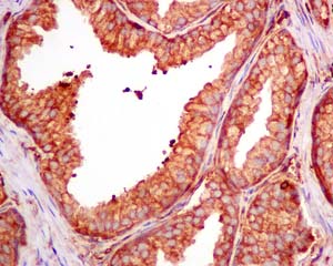
Immunohistochemical analysis of paraffin-embedded prostate tissue, labeling CAP1 with ab155079 at 1/100 dilution.
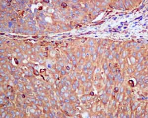
Immunohistochemical analysis of paraffin embedded Human cervical carcinoma tissue using ab155079 showing +ve staining.
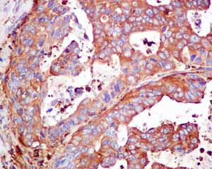
Immunohistochemical analysis of paraffin embedded Human gastric adenocarcinoma tissue using ab155079 showing +ve staining.
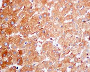
Immunohistochemical analysis of paraffin embedded Human normal liver tissue using ab155079 showing +ve staining.
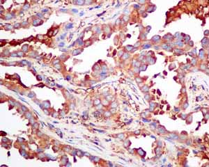
Immunohistochemical analysis of paraffin embedded Human lung adenocarcinoma tissue using ab155079 showing +ve staining.