![Anti-CCDC106 antibody (ab174943) at 1/100 dilution + Mouse cerebellum tissue lysate at 15 µg](http://www.bioprodhub.com/system/product_images/ab_products/2/sub_1/22206_ab174943-201045-ab174943.jpg)
Anti-CCDC106 antibody (ab174943) at 1/100 dilution + Mouse cerebellum tissue lysate at 15 µg
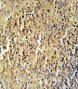
![Immunohistochemistry analysis of formalin-fixed and paraffin-embedded Human spleen tissue labeling CCDC106 using ab174943 at a 1/50 followed by peroxidase conjugation of the secondary antibody and DAB staining.](http://www.bioprodhub.com/system/product_images/ab_products/2/sub_1/22207_ab174943-201047-ab174943-2.jpg)
Immunohistochemistry analysis of formalin-fixed and paraffin-embedded Human spleen tissue labeling CCDC106 using ab174943 at a 1/50 followed by peroxidase conjugation of the secondary antibody and DAB staining.
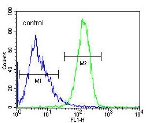
![Flow cytometric analysis of Jurkat cells (right histogram) compared to a negative control cell (left histogram) labeling CCDC106 using ab174943 at a 1/10 dilution. FITC-conjugated goat-anti-rabbit secondary antibodies were used for the analysis.](http://www.bioprodhub.com/system/product_images/ab_products/2/sub_1/22208_ab174943-201048-ab174943-3.jpg)
Flow cytometric analysis of Jurkat cells (right histogram) compared to a negative control cell (left histogram) labeling CCDC106 using ab174943 at a 1/10 dilution. FITC-conjugated goat-anti-rabbit secondary antibodies were used for the analysis.