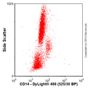
Human peripheral blood cells stained with ab36595 (red line). Human whole blood was processed using a modified protocol based on Chow et al, 2005 (PMID: 16080188). In brief, human whole blood was fixed in 4% formaldehyde (methanol-free) for 10 min at 22°C. Red blood cells were then lyzed by the addition of Triton X-100 (final concentration - 0.1%) for 15 min at 37°C. For experimentation, cells were treated with 50% methanol (-20°C) for 15 min at 4°C. Cells were then incubated with the antibody (ab36595, 0.01μg/1x106 cells) for 30 min at 4°C. The secondary antibody used was Alexa Fluor® 488 goat anti-mouse IgG (H&L) (ab150113) at 1/2000 dilution for 30 min at 4°C. Acquisition of >30,000 total events were collected using a 20mW Argon ion laser (488nm) and 525/30 bandpass filter.

Ab36595 at a 1/1300 dilution (Figure 1), and normal mouse serum at a 1/20 dilution (Figure 2), staining paraffin embedded human normal lymph node by immunohistochemistry. HRP-anti-mouse was used as the secondary antibody before colour development with DAB.

