![All lanes : Anti-CD166 antibody [EPR2759(2)] (ab109215) at 1/10000 dilution (purified)Lane 1 : SH-SY5Y cell lysateLane 2 : HuT-78 cell lysateLane 3 : HT-1080 cell lysateLysates/proteins at 20 µg per lane.SecondaryPeroxidase conjugated goat anti-rabbit IgG (H+L) at 1/1000 dilution](http://www.bioprodhub.com/system/product_images/ab_products/2/sub_1/23597_ab109215-238987-ab109215wb.jpg)
All lanes : Anti-CD166 antibody [EPR2759(2)] (ab109215) at 1/10000 dilution (purified)Lane 1 : SH-SY5Y cell lysateLane 2 : HuT-78 cell lysateLane 3 : HT-1080 cell lysateLysates/proteins at 20 µg per lane.SecondaryPeroxidase conjugated goat anti-rabbit IgG (H+L) at 1/1000 dilution
![All lanes : Anti-CD166 antibody [EPR2759(2)] (ab109215) at 1/10000 dilution (purified)Lane 1 : Mouse brain tissue lysateLane 2 : Rat brain tissue lysateLysates/proteins at 20 µg per lane.SecondaryPeroxidase conjugated goat anti-rabbit IgG (H+L) at 1/1000 dilution](http://www.bioprodhub.com/system/product_images/ab_products/2/sub_1/23598_ab109215-238988-ab109215wb2.jpg)
All lanes : Anti-CD166 antibody [EPR2759(2)] (ab109215) at 1/10000 dilution (purified)Lane 1 : Mouse brain tissue lysateLane 2 : Rat brain tissue lysateLysates/proteins at 20 µg per lane.SecondaryPeroxidase conjugated goat anti-rabbit IgG (H+L) at 1/1000 dilution
![All lanes : Anti-CD166 antibody [EPR2759(2)] (ab109215) at 1/1000 dilution (unpurified)Lane 1 : SH-SY5Y cell lysateLane 2 : HuT-78 cell lysateLane 3 : HT-1080 cell lysateLane 4 : Daudi cell lysateLysates/proteins at 10 µg per lane.SecondaryHRP labelled goat anti-rabbit at 1/2000 dilution](http://www.bioprodhub.com/system/product_images/ab_products/2/sub_1/23599_CD166-Primary-antibodies-ab109215-4.jpg)
All lanes : Anti-CD166 antibody [EPR2759(2)] (ab109215) at 1/1000 dilution (unpurified)Lane 1 : SH-SY5Y cell lysateLane 2 : HuT-78 cell lysateLane 3 : HT-1080 cell lysateLane 4 : Daudi cell lysateLysates/proteins at 10 µg per lane.SecondaryHRP labelled goat anti-rabbit at 1/2000 dilution
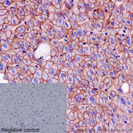
Immunohistochemistry (Formalin/PFA-fixed paraffin-embedded sections) analysis of human liver tissue labelling CD166 with purified ab109215 at 1/50. Heat mediated antigen retrieval was performed using Tris/EDTA buffer pH 9. A prediluted HRP-polymer conjugated anti-rabbit IgG was used as the secondary antibody. Negative control using PBS instead of primary antibody. Counterstained with hematoxylin.
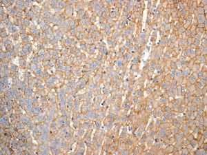
Immunohistochemistry (Formalin/PFA-fixed paraffin-embedded sections) analysis of human liver tissue labelling CD166 with unpurified ab109215 at 1/100.
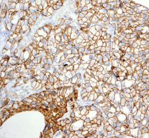
Immunohistochemistry (Formalin/PFA-fixed paraffin-embedded sections) analysis of human prostatic adenocarcinoma tissue labelling CD166 with unpurified ab109215 at 1/100.
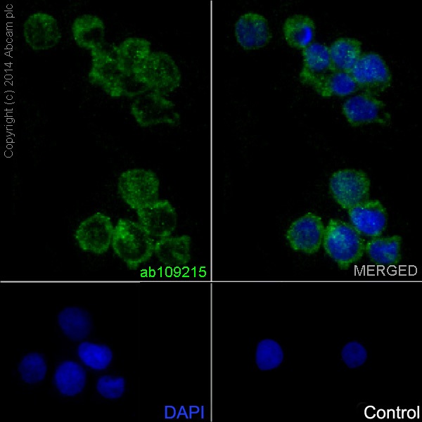
Immunocytochemistry/Immunofluorescence analysis of Jurkat cells labelling Cd166 (green) with purified ab109215 at 1/50. Cells were fixed with 4% paraformaldehyde and permeabilized with 0.1% Triton X-100. ab150077, an Alexa Fluor® 488-conjugated goat anti-rabbit IgG (1/500) was used as the secondary antibody. DAPI (blue) was used as the nuclear counterstain.Control: primary antibody (1/50) and secondary antibody ab150120, an Alexa Fluor® 594-conjugated goat anti-mouse IgG (1/500).
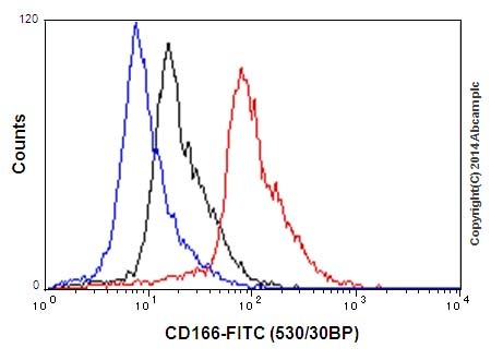
Flow Cytometry analysis of HuT-78 cells labelling CD166 with purified ab109215 at 1/90 (red). Cells were fixed with 2% paraformaldehyde. A FITC-conjugated goat anti-rabbit IgG (1/150) was used as the secondary antibody. Black - Isotype control, rabbit monoclonal IgG. Blue - Unlabelled control, cells without incubation with primary and secondary antibodies.
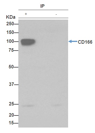
ab109215 (purified) at 1/30 immunoprecipitating CD166 in SH-SY5Y cell lysate (Lane 1). Lane 2 - PBS. For western blotting, a HRP-conjugated anti-rabbit IgG, specific to the non-reduced form of IgG was used as the secondary antibody (1/1500).Blocking buffer and concentration: 5% NFDM/TBST.Diluting buffer and concentration: 5% NFDM /TBST.
![All lanes : Anti-CD166 antibody [EPR2759(2)] (ab109215) at 1/10000 dilution (purified)Lane 1 : SH-SY5Y cell lysateLane 2 : HuT-78 cell lysateLane 3 : HT-1080 cell lysateLysates/proteins at 20 µg per lane.SecondaryPeroxidase conjugated goat anti-rabbit IgG (H+L) at 1/1000 dilution](http://www.bioprodhub.com/system/product_images/ab_products/2/sub_1/23597_ab109215-238987-ab109215wb.jpg)
![All lanes : Anti-CD166 antibody [EPR2759(2)] (ab109215) at 1/10000 dilution (purified)Lane 1 : Mouse brain tissue lysateLane 2 : Rat brain tissue lysateLysates/proteins at 20 µg per lane.SecondaryPeroxidase conjugated goat anti-rabbit IgG (H+L) at 1/1000 dilution](http://www.bioprodhub.com/system/product_images/ab_products/2/sub_1/23598_ab109215-238988-ab109215wb2.jpg)
![All lanes : Anti-CD166 antibody [EPR2759(2)] (ab109215) at 1/1000 dilution (unpurified)Lane 1 : SH-SY5Y cell lysateLane 2 : HuT-78 cell lysateLane 3 : HT-1080 cell lysateLane 4 : Daudi cell lysateLysates/proteins at 10 µg per lane.SecondaryHRP labelled goat anti-rabbit at 1/2000 dilution](http://www.bioprodhub.com/system/product_images/ab_products/2/sub_1/23599_CD166-Primary-antibodies-ab109215-4.jpg)


