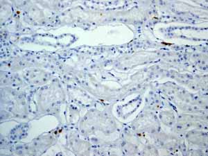
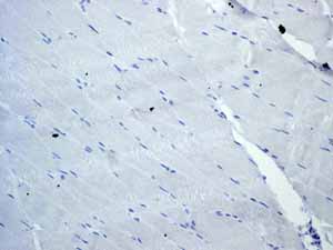
Anti-CD3 epsilon antibody [EP449E]
| Name | Anti-CD3 epsilon antibody [EP449E] |
|---|---|
| Supplier | Abcam |
| Catalog | ab52959 |
| Prices | $385.00 |
| Sizes | 100 µl |
| Host | Rabbit |
| Clonality | Monoclonal |
| Isotype | IgG |
| Clone | EP449E |
| Applications | WB IHC-P IP FC |
| Species Reactivities | Human |
| Antigen | A synthetic peptide corresponding to residues in cytoplasmic domain of human CD3 epsilon |
| Description | Rabbit Monoclonal |
| Gene | CD3E |
| Conjugate | Unconjugated |
| Supplier Page | Shop |
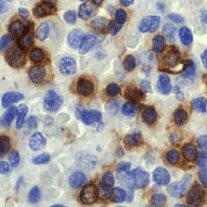
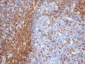
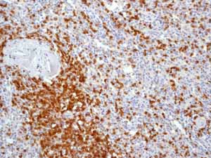
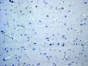
Product images
Product References
In situ characterization of intrahepatic non-parenchymal cells in PSC reveals - In situ characterization of intrahepatic non-parenchymal cells in PSC reveals
Berglin L, Bergquist A, Johansson H, Glaumann H, Jorns C, Lunemann S, Wedemeyer H, Ellis EC, Bjorkstrom NK. PLoS One. 2014 Aug 20;9(8):e105375.
Strong expression of TGF-beta in human host tissues around subcutaneous - Strong expression of TGF-beta in human host tissues around subcutaneous
Brattig NW, Racz P, Hoerauf A, Buttner DW. Parasitol Res. 2011 Jun;108(6):1347-54.
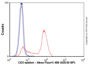
![Anti-CD3 epsilon antibody [EP449E] (ab52959) at 1/10000000 dilution + Jurkat cell lysate at 10 µgSecondarygoat anti-rabbit HRP at 1/2000 dilution](http://www.bioprodhub.com/system/product_images/ab_products/2/sub_1/24304_ab52959wb.gif)
