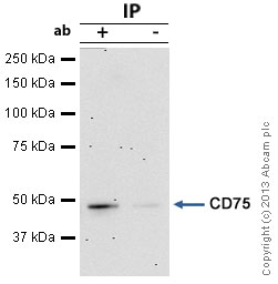
CD75 was immunoprecipitated using 0.5mg HepG2 whole cell extract, 5µg of Mouse monoclonal to CD75 and 50µl of protein G magnetic beads (+). No antibody was added to the control (-).The antibody was incubated under agitation with Protein G beads for 10min, HepG2 whole cell extract lysate diluted in RIPA buffer was added to each sample and incubated for a further 10min under agitation.Proteins were eluted by addition of 40µl SDS loading buffer and incubated for 10min at 70°C; 10µl of each sample was separated on a SDS PAGE gel, transferred to a nitrocellulose membrane, blocked with 5% BSA and probed with ab27316.Secondary: Goat polyclonal to mouse IgG light chain specific (HRP) at 1/20,000 dilution.Band: 47kDa; CD75
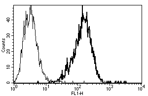
Ab27316, at a dilution of 10µl for 106 cells or 100 µl of whole blood, staining CD75 by Flow Cytometry.