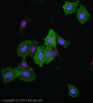
ICC/IF image of ab2528 stained MCF7 cells. The cells were 4% formaldehyde fixed (10 min) and then incubated in 1%BSA / 10% normal goat serum / 0.3M glycine in 0.1% PBS-Tween for 1h to permeabilise the cells and block non-specific protein-protein interactions. The cells were then incubated with the antibody (ab2528, 5µg/ml) overnight at +4°C. The secondary antibody (green) was ab96879, DyLight® 488 goat anti-mouse IgG (H+L) used at a 1/250 dilution for 1h. Alexa Fluor® 594 WGA was used to label plasma membranes (red) at a 1/200 dilution for 1h. DAPI was used to stain the cell nuclei (blue) at a concentration of 1.43µM.
![All lanes : Anti-CD98 antibody [MEM-108] (ab2528) at 5 µg/mlLane 1 : Jurkat (Human T cell lymphoblast-like cell line) Whole Cell Lysate Lane 2 : MCF7 (Human breast adenocarcinoma cell line) Whole Cell LysateLysates/proteins at 10 µg per lane.SecondaryGoat polyclonal to Mouse IgG - H&L - Pre-Adsorbed (HRP) at 1/3000 dilutionThe CD98 protein has a predicted molecular weight of 57 kDa, however it is extensively glycosylated. This antibody recognises a bands at 98-150 kDa, similar to what has been observed with other antibodies directed against this protein. Abcam welcomes customer feedback and would appreciate any comments regarding this product and the data presented above.](http://www.bioprodhub.com/system/product_images/ab_products/2/sub_1/26109_CD98-Primary-antibodies-ab2528-2.jpg)
All lanes : Anti-CD98 antibody [MEM-108] (ab2528) at 5 µg/mlLane 1 : Jurkat (Human T cell lymphoblast-like cell line) Whole Cell Lysate Lane 2 : MCF7 (Human breast adenocarcinoma cell line) Whole Cell LysateLysates/proteins at 10 µg per lane.SecondaryGoat polyclonal to Mouse IgG - H&L - Pre-Adsorbed (HRP) at 1/3000 dilutionThe CD98 protein has a predicted molecular weight of 57 kDa, however it is extensively glycosylated. This antibody recognises a bands at 98-150 kDa, similar to what has been observed with other antibodies directed against this protein. Abcam welcomes customer feedback and would appreciate any comments regarding this product and the data presented above.

![All lanes : Anti-CD98 antibody [MEM-108] (ab2528) at 5 µg/mlLane 1 : Jurkat (Human T cell lymphoblast-like cell line) Whole Cell Lysate Lane 2 : MCF7 (Human breast adenocarcinoma cell line) Whole Cell LysateLysates/proteins at 10 µg per lane.SecondaryGoat polyclonal to Mouse IgG - H&L - Pre-Adsorbed (HRP) at 1/3000 dilutionThe CD98 protein has a predicted molecular weight of 57 kDa, however it is extensively glycosylated. This antibody recognises a bands at 98-150 kDa, similar to what has been observed with other antibodies directed against this protein. Abcam welcomes customer feedback and would appreciate any comments regarding this product and the data presented above.](http://www.bioprodhub.com/system/product_images/ab_products/2/sub_1/26109_CD98-Primary-antibodies-ab2528-2.jpg)