Anti-CDRL2 antibody
| Name | Anti-CDRL2 antibody |
|---|---|
| Supplier | Abcam |
| Catalog | ab121705 |
| Prices | $443.00 |
| Sizes | 100 µl |
| Host | Rabbit |
| Clonality | Polyclonal |
| Isotype | IgG |
| Applications | ICC/IF ICC/IF WB IHC-P |
| Species Reactivities | Mouse, Rat, Human |
| Antigen | antigen sequence corresponding to amino acids 395-464 (DSSWRDLRGG EEGQGEVKAG EKSLSQHVEA VDKRLEQSQP EYKALFKEIF SRIQKTKADI NATKVKTHSS) of Human CDRL2 |
| Description | Rabbit Polyclonal |
| Gene | CDR2L |
| Conjugate | Unconjugated |
| Supplier Page | Shop |
Product images
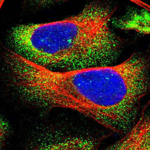 Immunofluorescent staining of Human cell line U-2 OS shows positivity in cytoplasm. Recommended concentration of ab121705 1-4 µg/ml. Cells treated with PFA/Triton X-100.
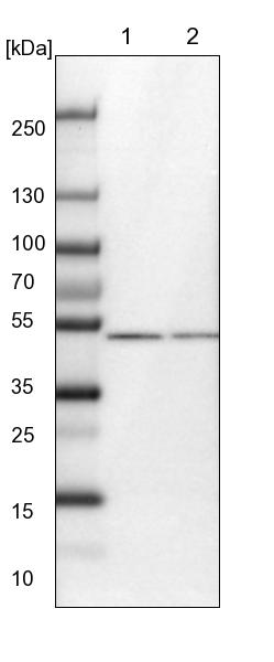 Lane 1: NIH-3T3 cell lysate (Mouse embryonic fibroblast cells)Lane 2: NBT-II cell lysate (Rat Wistar bladder tumour cells)
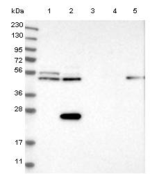 All lanes : Anti-CDRL2 antibody (ab121705) at 1/250 dilutionLane 1 : RT-4 cell lysate.Lane 2 : U-251 MG cell lysate.Lane 3 : Human PlasmaLane 4 : Human Liver tissue lysate.Lane 5 : Human Tonsil tissue lysate.developed using the ECL technique
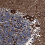 ab121705, at 1/125, staining CDRL2, in paraffin-embedded Human cerebellum tissue, by Immunohistochemistry.