Anti-CENPE antibody [1H12]
| Name | Anti-CENPE antibody [1H12] |
|---|---|
| Supplier | Abcam |
| Catalog | ab5093 |
| Prices | $401.00 |
| Sizes | 100 µl |
| Host | Mouse |
| Clonality | Monoclonal |
| Isotype | IgG1 |
| Clone | 1H12 |
| Applications | ICC/IF ICC/IF IP ICC/IF WB FC |
| Species Reactivities | Human |
| Antigen | Recombinant full length protein (Human) |
| Description | Mouse Monoclonal |
| Gene | CENPE |
| Conjugate | Unconjugated |
| Supplier Page | Shop |
Product images
Product References
CLIP-170 recruits PLK1 to kinetochores during early mitosis for chromosome - CLIP-170 recruits PLK1 to kinetochores during early mitosis for chromosome
Amin MA, Itoh G, Iemura K, Ikeda M, Tanaka K. J Cell Sci. 2014 Jul 1;127(Pt 13):2818-24.
MISP is a novel Plk1 substrate required for proper spindle orientation and - MISP is a novel Plk1 substrate required for proper spindle orientation and
Zhu M, Settele F, Kotak S, Sanchez-Pulido L, Ehret L, Ponting CP, Gonczy P, Hoffmann I. J Cell Biol. 2013 Mar 18;200(6):773-87.
Midbody assembly and its regulation during cytokinesis. - Midbody assembly and its regulation during cytokinesis.
Hu CK, Coughlin M, Mitchison TJ. Mol Biol Cell. 2012 Mar;23(6):1024-34.
Small GTPase Rab5 participates in chromosome congression and regulates - Small GTPase Rab5 participates in chromosome congression and regulates
Serio G, Margaria V, Jensen S, Oldani A, Bartek J, Bussolino F, Lanzetti L. Proc Natl Acad Sci U S A. 2011 Oct 18;108(42):17337-42. doi:
Aurora kinases and protein phosphatase 1 mediate chromosome congression through - Aurora kinases and protein phosphatase 1 mediate chromosome congression through
Kim Y, Holland AJ, Lan W, Cleveland DW. Cell. 2010 Aug 6;142(3):444-55.
Telomere disruption results in non-random formation of de novo dicentric - Telomere disruption results in non-random formation of de novo dicentric
Stimpson KM, Song IY, Jauch A, Holtgreve-Grez H, Hayden KE, Bridger JM, Sullivan BA. PLoS Genet. 2010 Aug 12;6(8). pii: e1001061.
Mediator of DNA damage checkpoint 1 (MDC1) regulates mitotic progression. - Mediator of DNA damage checkpoint 1 (MDC1) regulates mitotic progression.
Townsend K, Mason H, Blackford AN, Miller ES, Chapman JR, Sedgwick GG, Barone G, Turnell AS, Stewart GS. J Biol Chem. 2009 Dec 4;284(49):33939-48.
Glycogen synthase kinase 3beta interacts with and phosphorylates the - Glycogen synthase kinase 3beta interacts with and phosphorylates the
Cheng TS, Hsiao YL, Lin CC, Yu CT, Hsu CM, Chang MS, Lee CI, Huang CY, Howng SL, Hong YR. J Biol Chem. 2008 Jan 25;283(4):2454-64. Epub 2007 Nov 30.
Tankyrase-1 polymerization of poly(ADP-ribose) is required for spindle structure - Tankyrase-1 polymerization of poly(ADP-ribose) is required for spindle structure
Chang P, Coughlin M, Mitchison TJ. Nat Cell Biol. 2005 Nov;7(11):1133-9.
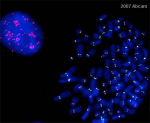
![Kinetochores specific staining of HCT116 cells arrested in G2/M phase by nocodazole treatment. Methanol fixed cells were stained using mouse monoclonal [1H12] antibody to CENP-E ab5093 (green) and DAPI (blue). This image was kindly supplied as part of the review submitted by Salvador Rodrigez-Nieto.](http://www.bioprodhub.com/system/product_images/ab_products/2/sub_1/27575_ab5093_1.jpg)


![All lanes : Anti-CENPE antibody [1H12] (ab5093) at 1 µg/mlLane 1 : HeLa (Human epithelial carcinoma cell line) Whole Cell LysateLane 2 : HepG2 (Human hepatocellular liver carcinoma cell line) Whole Cell Lysate Lane 3 : HEK293 (Human embryonic kidney cell line) Whole Cell LysateLysates/proteins at 10 µg per lane.SecondaryGoat Anti-Mouse IgG H&L (HRP) preadsorbed (ab97040) at 1/5000 dilutiondeveloped using the ECL techniquePerformed under reducing conditions.](http://www.bioprodhub.com/system/product_images/ab_products/2/sub_1/27578_CENPE-Primary-antibodies-ab5093-2.jpg)
![Overlay histogram showing HeLA cells stained with ab5093 (red line). The cells were fixed with 100% methanol (5 min) and then permeabilized with 0.1% PBS-Tween for 20 min. The cells were then incubated in 1x PBS / 10% normal goat serum / 0.3M glycine to block non-specific protein-protein interactions followed by the antibody (ab5093, 1µg/1x106 cells) for 30 min at 22°C. The secondary antibody used was DyLight® 488 goat anti-mouse IgG (H+L) (ab96879) at 1/500 dilution for 30 min at 22°C. Isotype control antibody (black line) was Mouse IgG1 [ICIGG1] (ab91353, 2µg/1x106 cells) used under the same conditions. Acquisition of >5,000 events was performed.](http://www.bioprodhub.com/system/product_images/ab_products/2/sub_1/27579_CENPE-Primary-antibodies-ab5093-3.jpg)