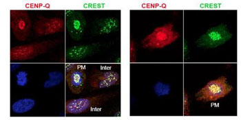
Immunofluorescence microscopy using ab105742 shows detection of endogenous CENPQ in HeLa whole cell lysate. Primary antibody was used at 1:100 followed by secondary antibody diluted 1:150. Red punctate anti-CENPQ signal colocalizes in overlay images with green punctate anti-CREST signals at the kinetochores (attached points of sister chromatids). Visible are colocalized CENPQ and CREST signal at various stages of the cell cycle as indicated from interphase to the end of mitosis. Nuclei are counter stained with bisbenzimide.

