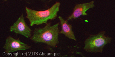
Immunocytochemistry/Immunofluorescence analysis of NBF-fixed asynchronous HeLa cells labelling CEP290 with ab85728 at 1/500. A DyLight® 594-conjugated goat anti-rabbit IgG (1/100) was used as the secondary antibody.

ICC/IF image of ab85728 stained HeLa cells. The cells were 4% formaldehyde fixed (10 min) and then incubated in 1%BSA / 10% normal goat serum / 0.3M glycine in 0.1% PBS-Tween for 1h to permeabilise the cells and block non-specific protein-protein interactions. The cells were then incubated with the antibody (ab85728, 5µg/ml) overnight at +4°C. The secondary antibody (green) was ab96899, DyLight® 488 goat anti-mouse IgG (H+L) used at a 1/250 dilution for 1h. Alexa Fluor® 594 WGA was used to label plasma membranes (red) at a 1/200 dilution for 1h. DAPI was used to stain the cell nuclei (blue) at a concentration of 1.43µM.

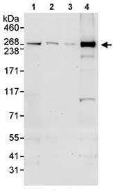
All lanes : Anti-CEP290 antibody (ab85728) at 0.1 µg/mlLane 1 : Whole cell lysate from HeLa cells at 50 µgLane 2 : Whole cell lysate from HeLa cells at 15 µgLane 3 : Whole cell lysate from HeLa cells at 5 µgLane 4 : Whole cell lysate from 293T cells at 50 µgdeveloped using the ECL technique

Detection of Human CEP290 in Immunoprecipitates of Whole cell lysate from HeLa cells (1 mg for IP, 20% of IP loaded) using ab85728 at 3 µg/mg lysate for IP (Lane 1) and at 1 µg/ml for subsequent Western blot detection. Lane 2 represents control IgG IP. Detection: Chemiluminescence with an exposure time of 10 seconds.


