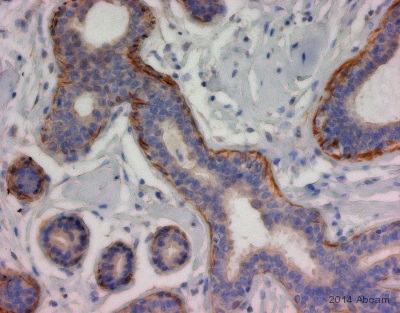
ab180914 staining CERD4 in human breast tissue sections by Immunohistochemistry (IHC-P - paraformaldehyde-fixed, paraffin-embedded sections). Tissue was fixed with paraformaldehyde and permeabilized with wash buffer + tween; antigen retrieval was by heat mediation in TRIS-EDTA buffer pH 9.0. Samples were incubated with primary antibody (1/500) for 30 minutes at 20°C. An undiluted HRP-conjugated goat anti-rabbit IgG polyclonal was used as the secondary antibody.See Abreview

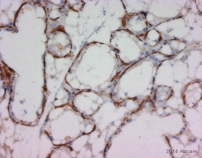
ab180914 staining CERD4 in mouse breast tissue sections by Immunohistochemistry (IHC-P - paraformaldehyde-fixed, paraffin-embedded sections). Tissue was fixed with paraformaldehyde and permeabilized with wash buffer + tween; antigen retrieval was by heat mediation in TRIS-EDTA buffer pH 9.0. Samples were incubated with primary antibody (1/500) for 30 minutes at 20°C. An undiluted HRP-conjugated goat anti-rabbit IgG polyclonal was used as the secondary antibody.See Abreview

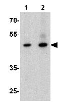
Lane 1 : Anti-CERD4 antibody - C-terminal (ab180914) at 1 µg/mlLane 2 : Anti-CERD4 antibody - C-terminal (ab180914) at 2 µg/mlLane 1 : Mouse brain tissue lysateLane 2 : Mouse brain tissue lysateLysates/proteins at 15 µg per lane.


