Anti-cIAP2 antibody
| Name | Anti-cIAP2 antibody |
|---|---|
| Supplier | Abcam |
| Catalog | ab23423 |
| Prices | $382.00 |
| Sizes | 100 µg |
| Host | Rabbit |
| Clonality | Polyclonal |
| Isotype | IgG |
| Applications | WB ICC/IF ICC/IF |
| Species Reactivities | Human, Mouse, Rat, Chicken, Dog |
| Antigen | Synthetic peptide conjugated to KLH derived from within residues 550 to the C-terminus of Human cIAP2 |
| Description | Rabbit Polyclonal |
| Gene | BIRC3 |
| Conjugate | Unconjugated |
| Supplier Page | Shop |
Product images
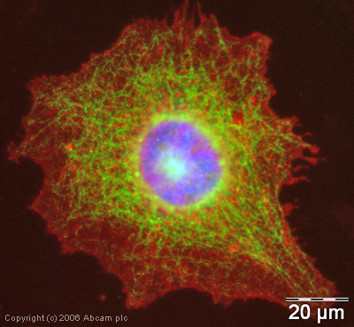 ICC/IF image of ab23423 stained human HeLa cells. The cells were methanol fixed (5 min) and incubated with the antibody (ab23423, 1µg/ml) for 1h at room temperature. The secondary antibody (green) was Alexa Fluor® 488 goat anti-rabbit IgG (H+L) used at a 1/1000 dilution for 1h. Image-iTTM FX Signal Enhancer was used as the primary blocking agent, 5% BSA (in TBS-T) was used for all other blocking steps. DAPI was used to stain the cell nuclei (blue). Alexa Fluor® 594 WGA was used to label plasma membranes (red).
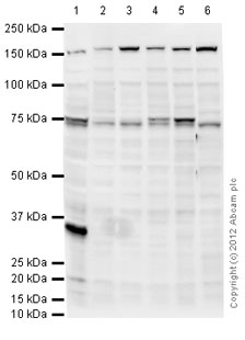 All lanes : Anti-cIAP2 antibody (ab23423) at 1 µg/mlLane 1 : Daudi (Human Burkitt's lymphoma cell line) Whole Cell LysateLane 2 : MOLT4 (Human acute lymphoblastic leukemia cell line) Whole Cell Lysate Lane 3 : Jurkat (Human T cell lymphoblast-like cell line) Whole Cell Lysate Lane 4 : Ramos (Human Burkitt's lymphoma cell line) Whole Cell Lysate Lane 5 : Raji (Human Burkitt's lymphoma cell line) Whole Cell LysateLane 6 : HepG2 (Human hepatocellular liver carcinoma cell line) Whole Cell Lysate Lysates/proteins at 10 µg/ml per lane.SecondaryGoat Anti-Rabbit IgG H&L (HRP) preadsorbed (ab97080) at 1/5000 dilutiondeveloped using the ECL techniquePerformed under reducing conditions.
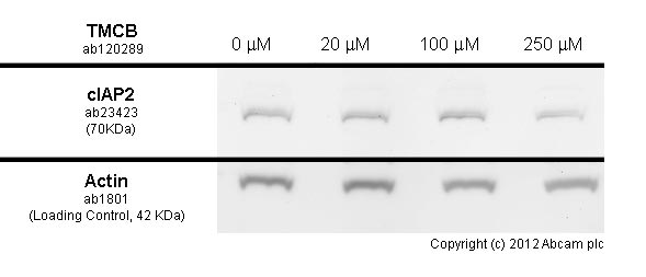
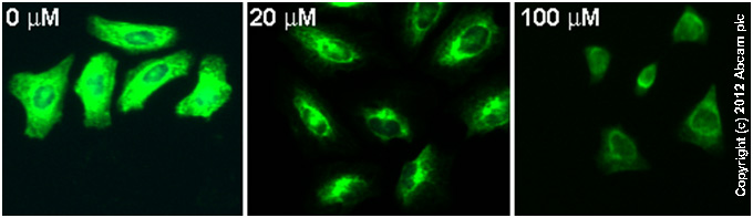 ab23423 staining cIAP2 in HeLa cells treated with TMCB (ab120289), by ICC/IF. Decrease in cIAP2 expression correlates with increased concentration of TMCB, as described in literature.The cells were incubated at 37°C for 10 minutes in media containing different concentrations of ab120289 (TMCB) in DMSO, fixed with 4% formaldehyde for 10 minutes at room temperature and blocked with PBS containing 10% goat serum, 0.3 M glycine, 1% BSA and 0.1% tween for 2h at room temperature. Staining of the treated cells with ab23423 (5 µg/ml) was performed overnight at 4°C in PBS containing 1% BSA and 0.1% tween. A DyLight 488 goat anti-rabbit polyclonal antibody (ab96899) at 1/250 dilution was used as the secondary antibody. Nuclei were counterstained with DAPI and are shown in blue.
 developed using the ECL techniquePerformed under reducing conditions.
Product References
Opposing effect of EGFRWT on EGFRvIII-mediated NF-kappaB activation with RIP1 as - Opposing effect of EGFRWT on EGFRvIII-mediated NF-kappaB activation with RIP1 as
Puliyappadamba VT, Chakraborty S, Chauncey SS, Li L, Hatanpaa KJ, Mickey B, Noorani S, Shu HK, Burma S, Boothman DA, Habib AA. Cell Rep. 2013 Aug 29;4(4):764-75.
cIAP1 and cIAP2 limit macrophage necroptosis by inhibiting Rip1 and Rip3 - cIAP1 and cIAP2 limit macrophage necroptosis by inhibiting Rip1 and Rip3
McComb S, Cheung HH, Korneluk RG, Wang S, Krishnan L, Sad S. Cell Death Differ. 2012 Nov;19(11):1791-801.
UXT-V1 protects cells against TNF-induced apoptosis through modulating complex II - UXT-V1 protects cells against TNF-induced apoptosis through modulating complex II
Huang Y, Chen L, Zhou Y, Liu H, Yang J, Liu Z, Wang C. Mol Biol Cell. 2011 Apr 15;22(8):1389-97.
Inhibition of hepatitis B virus replication by cIAP2 involves accelerating the - Inhibition of hepatitis B virus replication by cIAP2 involves accelerating the
Wang Z, Ni J, Li J, Shi B, Xu Y, Yuan Z. J Virol. 2011 Nov;85(21):11457-67.
Identification of a common subnuclear localization signal. - Identification of a common subnuclear localization signal.
Mekhail K, Rivero-Lopez L, Al-Masri A, Brandon C, Khacho M, Lee S. Mol Biol Cell. 2007 Oct;18(10):3966-77. Epub 2007 Jul 25.