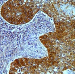
Immunohistochemical analysis of formalin-fixed, paraffin-embedded Human normal stomach tissue labeling Claudin18 with ab203563 at 2 µg/mL followed by DAB staining. 40x magnification.

Immunohistochemical analysis of formalin-fixed, paraffin-embedded Human squamous lung carcinoma tissue labeling Claudin18 with ab203563 at 2 µg/mL followed by DAB staining. 20x magnification.
![All lanes : Anti-Claudin18 antibody [34H14L15] (ab203563) at 2 µg/mlLane 1 : mouse lung lysateLane 2 : mouse lung lysate with immunizing peptide](http://www.bioprodhub.com/system/product_images/ab_products/2/sub_1/29448_ab203563-245492-ab2035633.jpg)
All lanes : Anti-Claudin18 antibody [34H14L15] (ab203563) at 2 µg/mlLane 1 : mouse lung lysateLane 2 : mouse lung lysate with immunizing peptide

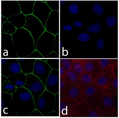
Immunofluorescent analysis of Caco-2 cells (4% paraformaldehyde-fixed) labeling Claudin18 with ab203563 at 1/500 dilution followed with Alexa Flour® 488 goat anti-rabbit IgG secondary antibody at 1/400 dilution (Panel a: green). Nuclei (Panel b: blue) were stained with DAPI. Panel c is a merged image showing cell junction localization and panel d is a control without primary antibody. 20X magnification.
![All lanes : Anti-Claudin18 antibody [34H14L15] (ab203563) at 1/1000 dilutionLane 1 : Rat heart lysateLane 2 : A431 lysateLane 3 : A549 lysateLysates/proteins at 30 µg per lane.Secondarygoat anti-rabbit HRP at 1/5000 dilution](http://www.bioprodhub.com/system/product_images/ab_products/2/sub_1/29450_ab203563-245490-ab2035635.jpg)
All lanes : Anti-Claudin18 antibody [34H14L15] (ab203563) at 1/1000 dilutionLane 1 : Rat heart lysateLane 2 : A431 lysateLane 3 : A549 lysateLysates/proteins at 30 µg per lane.Secondarygoat anti-rabbit HRP at 1/5000 dilution


![All lanes : Anti-Claudin18 antibody [34H14L15] (ab203563) at 2 µg/mlLane 1 : mouse lung lysateLane 2 : mouse lung lysate with immunizing peptide](http://www.bioprodhub.com/system/product_images/ab_products/2/sub_1/29448_ab203563-245492-ab2035633.jpg)

![All lanes : Anti-Claudin18 antibody [34H14L15] (ab203563) at 1/1000 dilutionLane 1 : Rat heart lysateLane 2 : A431 lysateLane 3 : A549 lysateLysates/proteins at 30 µg per lane.Secondarygoat anti-rabbit HRP at 1/5000 dilution](http://www.bioprodhub.com/system/product_images/ab_products/2/sub_1/29450_ab203563-245490-ab2035635.jpg)