Anti-Collagen I antibody
| Name | Anti-Collagen I antibody |
|---|---|
| Supplier | Abcam |
| Catalog | ab34710 |
| Prices | $403.00 |
| Sizes | 100 µg |
| Host | Rabbit |
| Clonality | Polyclonal |
| Isotype | IgG |
| Applications | IHC-F ELISA WB ELISA IHC-P ICC/IF ICC/IF IP |
| Species Reactivities | Mouse, Rat, Goat, Horse, Bovine, Human, Pig |
| Antigen | Full length native protein (purified) corresponding to Human Collagen I aa 1-1464 |
| Description | Rabbit Polyclonal |
| Gene | COL1A1 |
| Conjugate | Unconjugated |
| Supplier Page | Shop |
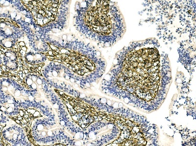
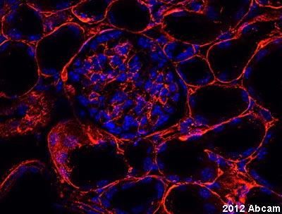
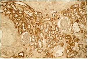
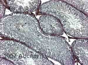
Product images
Product References
Endothelial cell microRNA expression in human late-onset Fuchs' dystrophy. - Endothelial cell microRNA expression in human late-onset Fuchs' dystrophy.
Matthaei M, Hu J, Kallay L, Eberhart CG, Cursiefen C, Qian J, Lackner EM, Jun AS. Invest Ophthalmol Vis Sci. 2014 Jan 9;55(1):216-25.
Mosquito saliva serine protease enhances dissemination of dengue virus into the - Mosquito saliva serine protease enhances dissemination of dengue virus into the
Conway MJ, Watson AM, Colpitts TM, Dragovic SM, Li Z, Wang P, Feitosa F, Shepherd DT, Ryman KD, Klimstra WB, Anderson JF, Fikrig E. J Virol. 2014 Jan;88(1):164-75.
Dystrophic changes in extraocular muscles after gamma irradiation in - Dystrophic changes in extraocular muscles after gamma irradiation in
McDonald AA, Kunz MD, McLoon LK. PLoS One. 2014 Jan 21;9(1):e86424.
Exaggerated renal fibrosis in P2X4 receptor-deficient mice following unilateral - Exaggerated renal fibrosis in P2X4 receptor-deficient mice following unilateral
Kim MJ, Turner CM, Hewitt R, Smith J, Bhangal G, Pusey CD, Unwin RJ, Tam FW. Nephrol Dial Transplant. 2014 Jul;29(7):1350-61.
Experimental orthotopic transplantation of a tissue-engineered oesophagus in - Experimental orthotopic transplantation of a tissue-engineered oesophagus in
Sjoqvist S, Jungebluth P, Lim ML, Haag JC, Gustafsson Y, Lemon G, Baiguera S, Burguillos MA, Del Gaudio C, Rodriguez AB, Sotnichenko A, Kublickiene K, Ullman H, Kielstein H, Damberg P, Bianco A, Heuchel R, Zhao Y, Ribatti D, Ibarra C, Joseph B, Taylor DA, Macchiarini P. Nat Commun. 2014 Apr 15;5:3562.
Diabetes increases pancreatic fibrosis during chronic inflammation. - Diabetes increases pancreatic fibrosis during chronic inflammation.
Zechner D, Knapp N, Bobrowski A, Radecke T, Genz B, Vollmar B. Exp Biol Med (Maywood). 2014 Jun;239(6):670-6.
The requirement of CD8+ T cells to initiate and augment acute cardiac - The requirement of CD8+ T cells to initiate and augment acute cardiac
Ma F, Feng J, Zhang C, Li Y, Qi G, Li H, Wu Y, Fu Y, Zhao Y, Chen H, Du J, Tang H. J Immunol. 2014 Apr 1;192(7):3365-73.
Haploinsufficiency of Def activates p53-dependent TGFbeta signalling and causes - Haploinsufficiency of Def activates p53-dependent TGFbeta signalling and causes
Zhu Z, Chen J, Xiong JW, Peng J. PLoS One. 2014 May 6;9(5):e96576.
Combinatorial scaffold morphologies for zonal articular cartilage engineering. - Combinatorial scaffold morphologies for zonal articular cartilage engineering.
Steele JA, McCullen SD, Callanan A, Autefage H, Accardi MA, Dini D, Stevens MM. Acta Biomater. 2014 May;10(5):2065-75.
Chronic hypoxia-inducible transcription factor-2 activation stably transforms - Chronic hypoxia-inducible transcription factor-2 activation stably transforms
Kurt B, Gerl K, Karger C, Schwarzensteiner I, Kurtz A. J Am Soc Nephrol. 2015 Mar;26(3):587-96.
