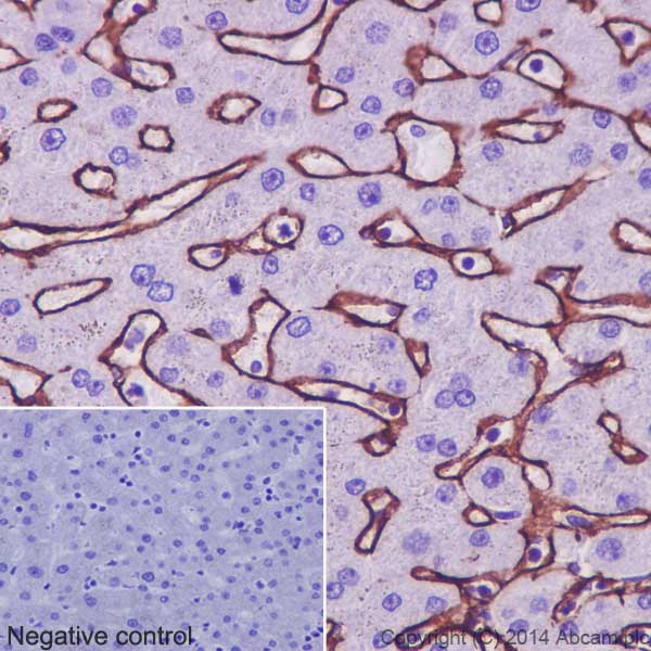
Immunohistochemical analysis of paraffin-embedded Human liver tissue labeling Collagen VI with ab182744 at 1/250 dilution, followed by prediluted HRP Polymer for Rabbit/Mouse IgG. Positive staining around sinusoidal endothelial basement membranes is observed. Counter stained with Hematoxylin.Negative control: Using PBS instead of primary ab, secondary ab is prediluted HRP Polymer for Rabbit/Mouse IgG.
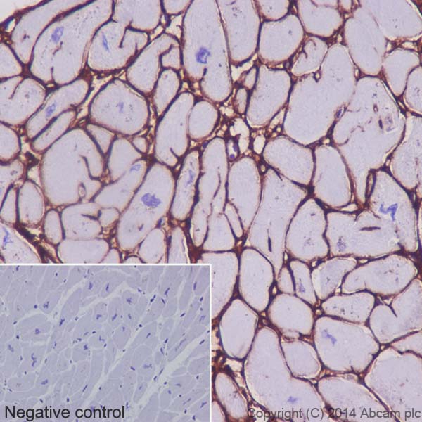
Immunohistochemical analysis of paraffin-embedded Human cardiac muscle tissue labeling Collagen VI with ab182744 at 1/250 dilution, followed by prediluted HRP Polymer for Rabbit/Mouse IgG. Positive staining on Human cardiac sarcolemma and interstitium is observed. Counter stained with Hematoxylin.Negative control: Using PBS instead of primary ab, secondary ab is prediluted HRP Polymer for Rabbit/Mouse IgG.
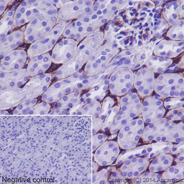
Immunohistochemical analysis of paraffin-embedded Mouse kidney tissue labeling Collagen VI with ab182744 at 1/250 dilution, followed by prediluted HRP Polymer for Rabbit/Mouse IgG. Positive staining around basement membranes of Mouse renal tubules is observed. Counter stained with Hematoxylin.Negative control: Using PBS instead of primary ab, secondary ab is prediluted HRP Polymer for Rabbit/Mouse IgG.
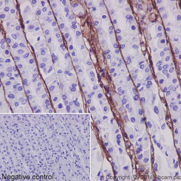
Immunohistochemical analysis of paraffin-embedded Rat stomach tissue labeling Collagen VI with ab182744 at 1/250 dilution, followed by prediluted HRP Polymer for Rabbit/Mouse IgG. Positive staining around Rat gastric epithelial basement membranes is observed. Counter stained with Hematoxylin.Negative control: Using PBS instead of primary ab, secondary ab is prediluted HRP Polymer for Rabbit/Mouse IgG.
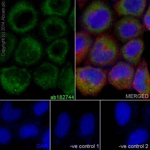
Immunofluorescent analysis of 4% paraformaldehyde-fixed, 0.1% Triton X-100 permeabilized HeLa (Human epithelial cells from cervix adenocarcinoma) cells labeling Collagen VI with ab182744 at 1/200 dilution, followed by Goat anti-rabbit IgG (Alexa Fluor® 488) (ab150077) secondary antibody at 1/400 dilution (green). Cytoplasm staining on HeLa cell line is observed. The nuclear counter stain is DAPI (blue). Tubulin is detected with ab7291 (anti-Tubulin mouse mAb) at 1/500 dilution and ab150120 (AlexaFluor®594 Goat anti-Mouse secondary) at 1/500 dilution (red).The negative controls are as follows:--ve control1 - ab182744 at 1/200 dilution followed by ab150120 (AlexaFluor®594 Goat anti-Mouse secondary) at 1/500 dilution.-ve control 2 - ab7291 (anti-Tubulin mouse mAb) at 1/500 dilution followed by ab150077 (Alexa Fluor®488 Goat Anti-Rabbit IgG H&L) at 1/400 dilution.
![All lanes : Anti-Collagen VI antibody [EPR17072] (ab182744) at 1/20000 dilutionLane 1 : Human skeletal muscle lysateLane 2 : WI-38 (Human fetal lung fibroblast cells) whole cell lysateLane 3 : Human placenta lysateLysates/proteins at 20 µg per lane.SecondaryAnti-Rabbit IgG (HRP), specific to the non-reduced form of IgG at 1/1000 dilution](http://www.bioprodhub.com/system/product_images/ab_products/2/sub_2/635_ab182744-231121-CollagenVIWB1.jpg)
All lanes : Anti-Collagen VI antibody [EPR17072] (ab182744) at 1/20000 dilutionLane 1 : Human skeletal muscle lysateLane 2 : WI-38 (Human fetal lung fibroblast cells) whole cell lysateLane 3 : Human placenta lysateLysates/proteins at 20 µg per lane.SecondaryAnti-Rabbit IgG (HRP), specific to the non-reduced form of IgG at 1/1000 dilution
![All lanes : Anti-Collagen VI antibody [EPR17072] (ab182744) at 1/2000 dilutionLane 1 : Human fetal brain lysateLane 2 : Human fetal heart lysateLane 3 : Human fetal kidney lysateLane 4 : Human fetal spleen lysateLysates/proteins at 10 µg per lane.SecondaryAnti-Rabbit IgG (HRP), specific to the non-reduced form of IgG at 1/1000 dilution](http://www.bioprodhub.com/system/product_images/ab_products/2/sub_2/636_ab182744-231122-CollagenVIWB2.jpg)
All lanes : Anti-Collagen VI antibody [EPR17072] (ab182744) at 1/2000 dilutionLane 1 : Human fetal brain lysateLane 2 : Human fetal heart lysateLane 3 : Human fetal kidney lysateLane 4 : Human fetal spleen lysateLysates/proteins at 10 µg per lane.SecondaryAnti-Rabbit IgG (HRP), specific to the non-reduced form of IgG at 1/1000 dilution
![All lanes : Anti-Collagen VI antibody [EPR17072] (ab182744) at 1/2000 dilutionLane 1 : Mouse heart lysateLane 2 : Mouse kidney lysateLane 3 : Mouse spleen lysateLane 4 : Rat kidney lysateLane 5 : Rat spleen lysateLane 6 : NIH/3T3 (Mouse embyro fibroblast cells) whole cell lysateLysates/proteins at 10 µg per lane.SecondaryGoat Anti-Rabbit IgG, (H+L),Peroxidase conjugated at 1/1000 dilution](http://www.bioprodhub.com/system/product_images/ab_products/2/sub_2/637_ab182744-231123-CollagenVIWB3.jpg)
All lanes : Anti-Collagen VI antibody [EPR17072] (ab182744) at 1/2000 dilutionLane 1 : Mouse heart lysateLane 2 : Mouse kidney lysateLane 3 : Mouse spleen lysateLane 4 : Rat kidney lysateLane 5 : Rat spleen lysateLane 6 : NIH/3T3 (Mouse embyro fibroblast cells) whole cell lysateLysates/proteins at 10 µg per lane.SecondaryGoat Anti-Rabbit IgG, (H+L),Peroxidase conjugated at 1/1000 dilution