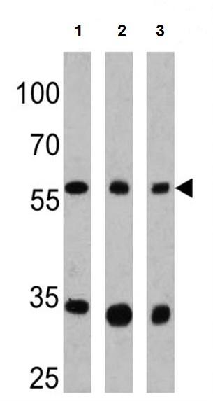
All lanes : Anti-CPEB1 antibody (ab3465) at 1/500 dilutionLane 1 : HeLa lysateLane 2 : Human brain tissue lysateLane 3 : Mouse brain tissue lysateLysates/proteins at 25 µg per lane.SecondaryHRP-conjugated anti-rabbitdeveloped using the ECL technique

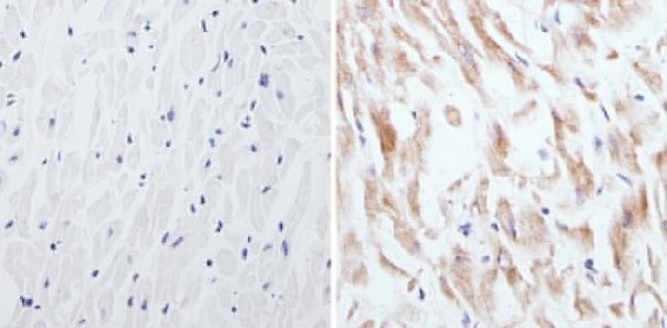
Immunohistochemistry analysis of CPEB showing staining in the cytoplasm of paraffin-embedded human hearat tissue (right) compared to a negative control without primary antibody (left). To expose target proteins, antigen retrieval was performed using 10 mM sodium citrate (pH 6.0), microwaved for 8-15 min. Following antigen retrieval, tissues were blocked in 3% H2O2-methanol for 15 min at room temperature, washed with ddH2O and PBS, and then probed with ab3465 diluted in 3% BSA-PBS at a dilution of 1/50 overnight at 4°C in a humidified chamber. Tissues were washed extensively in PBST and detection was performed using an HRP-conjugated secondary antibody followed by colorimetric detection using a DAB kit. Tissues were counterstained with hematoxylin and dehydrated with ethanol and xylene to prep for mounting.
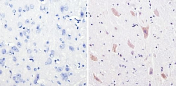
Immunohistochemistry analysis of CPEB showing staining in the cytoplasm of paraffin-embedded mouse brain tissue (right) compared to a negative control without primary antibody (left). To expose target proteins, antigen retrieval was performed using 10 mM sodium citrate (pH 6.0), microwaved for 8-15 min. Following antigen retrieval, tissues were blocked in 3% H2O2-methanol for 15 min at room temperature, washed with ddH2O and PBS, and then probed with ab3465 diluted in 3% BSA-PBS at a dilution of 1/200 overnight at 4°C in a humidified chamber. Tissues were washed extensively in PBST and detection was performed using an HRP-conjugated secondary antibody followed by colorimetric detection using a DAB kit. Tissues were counterstained with hematoxylin and dehydrated with ethanol and xylene to prep for mounting.
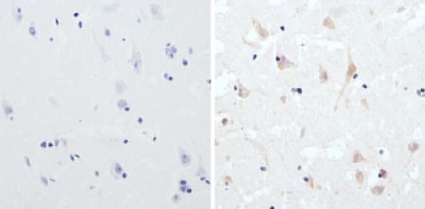
Immunohistochemistry analysis of CPEB showing staining in the cytoplasm of paraffin-embedded human brain tissue (right) compared to a negative control without primary antibody (left). To expose target proteins, antigen retrieval was performed using 10 mM sodium citrate (pH 6.0), microwaved for 8-15 min. Following antigen retrieval, tissues were blocked in 3% H2O2-methanol for 15 min at room temperature, washed with ddH2O and PBS, and then probed with ab3465 diluted in 3% BSA-PBS at a dilution of 1/100 overnight at 4°C in a humidified chamber. Tissues were washed extensively in PBST and detection was performed using an HRP-conjugated secondary antibody followed by colorimetric detection using a DAB kit. Tissues were counterstained with hematoxylin and dehydrated with ethanol and xylene to prep for mounting.
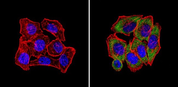
Immunofluorescent analysis of CPEB (green) showing staining in the cytoplasm of U251 cells (right) compared to a negative control without primary antibody (left). Formalin-fixed cells were permeabilized with 0.1% Triton X-100 in TBS for 5-10 minutes and blocked with 3% BSA-PBS for 30 minutes at room temperature. Cells were probed with ab3465 in 3% BSA-PBS at a dilution of 1/100 and incubated overnight at 4°C in a humidified chamber. Cells were washed with PBST and incubated with a DyLight®-conjugated secondary antibody in PBS at room temperature in the dark. Actin was stained using Alexa Fluor® 554 (red) and nuclei were stained with Hoechst or DAPI (blue). Images were taken at a magnification of 60x.
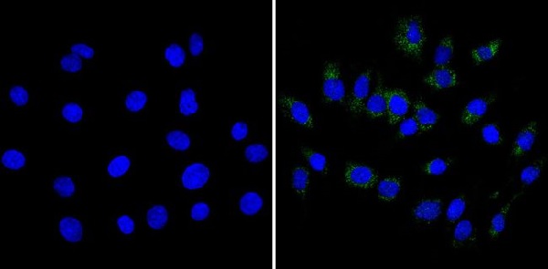
Immunofluorescent analysis of CPEB (green) showing staining in the cytoplasm of C6 cells (right) compared to a negative control without primary antibody (left). Formalin-fixed cells were permeabilized with 0.1% Triton X-100 in TBS for 5-10 minutes and blocked with 3% BSA-PBS for 30 minutes at room temperature. Cells were probed with ab3465 in 3% BSA-PBS at a dilution of 1/100 and incubated overnight at 4°C in a humidified chamber. Cells were washed with PBST and incubated with a DyLight®-conjugated secondary antibody in PBS at room temperature in the dark. Nuclei were stained with Hoechst or DAPI (blue). Images were taken at a magnification of 60x.
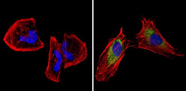
Immunofluorescent analysis of CPEB (green) showing staining in the cytoplasm of HeLa cells (right) compared to a negative control without primary antibody (left). Formalin-fixed cells were permeabilized with 0.1% Triton X-100 in TBS for 5-10 minutes and blocked with 3% BSA-PBS for 30 minutes at room temperature. Cells were probed with ab3465 in 3% BSA-PBS at a dilution of 1/100 and incubated overnight at 4°C in a humidified chamber. Cells were washed with PBST and incubated with a DyLight®-conjugated secondary antibody in PBS at room temperature in the dark. Actin was stained using Alexa Fluor® 554 (red) and nuclei were stained with Hoechst or DAPI (blue). Images were taken at a magnification of 60x.


