![All lanes : Anti-CSN1 antibody [EPR15642] (ab187658) at 1/10000 dilutionLane 1 : K562 cell lysateLane 2 : HeLa cell lysateLane 3 : MCF7 cell lysateLane 4 : 293T cell lysateLysates/proteins at 20 µg per lane.SecondaryGoat Anti-Rabbit IgG, (H+L), Peroxidase conjugated at 1/1000 dilution](http://www.bioprodhub.com/system/product_images/ab_products/2/sub_2/2780_ab187658-220696-ab1876581.jpg)
All lanes : Anti-CSN1 antibody [EPR15642] (ab187658) at 1/10000 dilutionLane 1 : K562 cell lysateLane 2 : HeLa cell lysateLane 3 : MCF7 cell lysateLane 4 : 293T cell lysateLysates/proteins at 20 µg per lane.SecondaryGoat Anti-Rabbit IgG, (H+L), Peroxidase conjugated at 1/1000 dilution
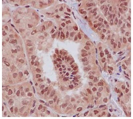
Immunohistochemical analysis of paraffin embedded Human thyroid papillary adenocarcinoma tissue labeling CSN1 with ab187658 at 1/250 dilution. Secondary staining with HRP Polymer for Rabbit IgG and counterstained with Hematoxylin.
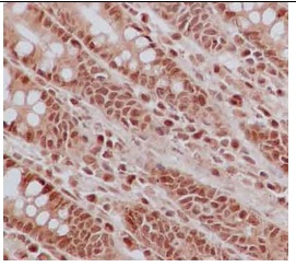
Immunohistochemical analysis of paraffin embedded Human colon tissue labeling CSN1 with ab187658 at 1/250 dilution. Secondary staining with HRP Polymer for Rabbit IgG and counterstained with Hematoxylin.
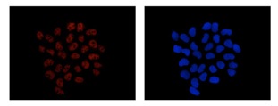
Immunofluorescent staining of 4% paraformaldehyde fixed HeLa cells labeling CSN1 with ab187658 at 1/250 dilution (red; left image) and DAPI nuclear staining (blue; right image). Secondary staining with Goat anti Rabbit IgG (Alexa Flour®555).
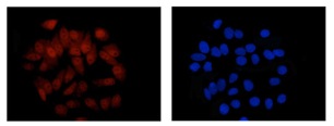
Immunofluorescent staining of 4% paraformaldehyde fixed MCF7 cells labeling CSN1 with ab187658 at 1/250 dilution (red; left image) and DAPI nuclear staining (blue; right image). Secondary staining with Goat anti Rabbit IgG (Alexa Flour®555).