All lanes : Anti-CtBP1 antibody (ab79417) at 1/500 dilutionLane 1 : Extracts from Jurkat cellsLane 2 : Extracts from Jurkat cells with immunizing peptide at 10 µgLysates/proteins at 30 µg per lane.

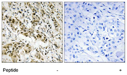
ab79417, at a 1/50 dilution, staining CtBP1 in paraffin embedded human breast carcinoma tissue by Immunohistochemistry in the absence (left panel) or presence (right panel) of the immunizing peptide.

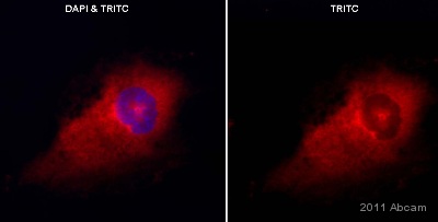
ab79417 staining CtBP1 in human glioblastoma cell line D54MG by Immunocytochemistry/ Immunofluorescence. Cells were fixed in paraformaldehyde and permeabilized in 0.1% Triton X-100 prior to blocking in 0.5% BSA for 20 minutes at room temperature. The primary antibody was diluted 1/60 in 0.5% BSA/PBS and incubated with the sample for 16 hours at 4°C. The secondary antibody was TRITC-conjugated Goat anti-Rabbit polyclonal, diluted 1/300.See Abreview

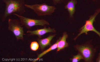
ICC/IF image of ab79417 stained HeLa cells. The cells were 4% formaldehyde (10 min) and then incubated in 1%BSA / 10% normal goat serum / 0.3M glycine in 0.1% PBS-Tween for 1h to permeabilise the cells and block non-specific protein-protein interactions. The cells were then incubated with the antibody (ab79417, 10µg/ml) overnight at +4°C. The secondary antibody (green) was for 1h. Alexa Fluor® 594 WGA was used to label plasma membranes (red) at a 1/200 dilution for 1h. DAPI was used to stain the cell nuclei (blue) at a concentration of 1.43µM.


