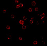
Anti-CXCR4 antibody
| Name | Anti-CXCR4 antibody |
|---|---|
| Supplier | Abcam |
| Catalog | ab2074 |
| Host | Rabbit |
| Clonality | Polyclonal |
| Isotype | IgG |
| Applications | FC IHC-P IHC-F IP WB ICC/IF ICC/IF |
| Species Reactivities | Mouse, Rat, Human |
| Antigen | Synthetic peptide, corresponding to N terminal amino acids 1-14 of Human CXCR4(Peptide available as ab8126 |
| Blocking Peptide | CXCR4 peptide |
| Description | Rabbit Polyclonal |
| Gene | CXCR4 |
| Conjugate | Unconjugated |
| Supplier Page | Shop |
Product images
Product References
Instruction of circulating endothelial progenitors in vitro towards specialized - Instruction of circulating endothelial progenitors in vitro towards specialized
Boyer-Di Ponio J, El-Ayoubi F, Glacial F, Ganeshamoorthy K, Driancourt C, Godet M, Perriere N, Guillevic O, Couraud PO, Uzan G. PLoS One. 2014 Jan 2;9(1):e84179.
CXCL12 chemokine expression suppresses human pancreatic cancer growth and - CXCL12 chemokine expression suppresses human pancreatic cancer growth and
Roy I, Zimmerman NP, Mackinnon AC, Tsai S, Evans DB, Dwinell MB. PLoS One. 2014 Mar 4;9(3):e90400.
CXCR4 inhibition ameliorates severe obliterative pulmonary hypertension and - CXCR4 inhibition ameliorates severe obliterative pulmonary hypertension and
Farkas D, Kraskauskas D, Drake JI, Alhussaini AA, Kraskauskiene V, Bogaard HJ, Cool CD, Voelkel NF, Farkas L. PLoS One. 2014 Feb 24;9(2):e89810.
The WIP1 oncogene promotes progression and invasion of aggressive medulloblastoma - The WIP1 oncogene promotes progression and invasion of aggressive medulloblastoma
Buss MC, Remke M, Lee J, Gandhi K, Schniederjan MJ, Kool M, Northcott PA, Pfister SM, Taylor MD, Castellino RC. Oncogene. 2015 Feb 26;34(9):1126-40.
The role of macrophage migration inhibitory factor in anesthetic-induced - The role of macrophage migration inhibitory factor in anesthetic-induced
Goetzenich A, Kraemer S, Rossaint R, Bleilevens C, Dollo F, Siry L, Rajabi-Alampour S, Beckers C, Soppert J, Lue H, Rex S, Bernhagen J, Stoppe C. PLoS One. 2014 Mar 25;9(3):e92827.
Silencing of CXCR4 inhibits tumor cell proliferation and neural invasion in human - Silencing of CXCR4 inhibits tumor cell proliferation and neural invasion in human
Tan XY, Chang S, Liu W, Tang HH. Gut Liver. 2014 Mar;8(2):196-204.
Gr-1+CD11b+ cells facilitate Lewis lung cancer recurrence by enhancing - Gr-1+CD11b+ cells facilitate Lewis lung cancer recurrence by enhancing
Liu T, Xie C, Ma H, Zhang S, Liang Y, Shi L, Yu D, Feng Y, Zhang T, Wu G. Sci Rep. 2014 Apr 29;4:4833.
CXCL12 in astrocytes contributes to bone cancer pain through CXCR4-mediated - CXCL12 in astrocytes contributes to bone cancer pain through CXCR4-mediated
Shen W, Hu XM, Liu YN, Han Y, Chen LP, Wang CC, Song C. J Neuroinflammation. 2014 Apr 16;11:75.
PIM kinases are essential for chronic lymphocytic leukemia cell survival (PIM2/3) - PIM kinases are essential for chronic lymphocytic leukemia cell survival (PIM2/3)
Decker S, Finter J, Forde AJ, Kissel S, Schwaller J, Mack TS, Kuhn A, Gray N, Follo M, Jumaa H, Burger M, Zirlik K, Pfeifer D, Miduturu CV, Eibel H, Veelken H, Dierks C. Mol Cancer Ther. 2014 May;13(5):1231-45.
Targeting CXCR7/ACKR3 as a therapeutic strategy to promote remyelination in the - Targeting CXCR7/ACKR3 as a therapeutic strategy to promote remyelination in the
Williams JL, Patel JR, Daniels BP, Klein RS. J Exp Med. 2014 May 5;211(5):791-9.


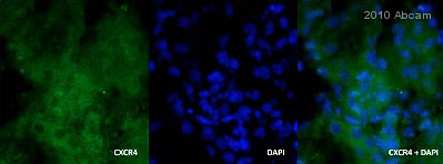
![ab2074 at 1/100 staining differentiated rat skeletal myocytes by ICC/IF. The cells were 2% paraformaldehyde fixed for 15 minutes and then incubated cells with ab2074 overnight at 4°C. The image demonstrates CXCR4 expressing skeletal myocytes (green-cytoplasmic and cell membrane localization) with DAPI (blue-nuclear counter stain) [Right panel] and DIC (phase) image of the same skeletal myocytes ([left panel].See Abreview](http://www.bioprodhub.com/system/product_images/ab_products/2/sub_2/3629_ab2074_3.jpg)


