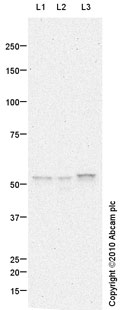
All lanes : Anti-Cyclin A antibody (ab87359) at 1 µg/mlLane 1 : F9 (Mouse embryonic carcinoma cell line) Whole Cell Lysate Lane 2 : MEF1 (Mouse embryonic fibroblast cell line) Whole Cell LysateLane 3 : K562 (Human erythromyeloblastoid leukemia cell line) Whole Cell Lysate Lysates/proteins at 10 µg per lane.SecondaryGoat polyclonal to Rabbit IgG - H&L - Pre-Adsorbed (HRP) at 1/3000 dilutiondeveloped using the ECL techniquePerformed under reducing conditions.
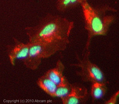
ICC/IF image of ab87359 stained HepG2 cells. The cells were 100% Methanol fixed (5 min) and then incubated in 1%BSA / 10% normal Goat serum / 0.3M glycine in 0.1% PBS-Tween for 1h to permeabilise the cells and block non-specific protein-protein interactions. The cells were then incubated with the antibody (ab87359, 5µg/ml) overnight at +4°C. The secondary antibody (green) was Alexa Fluor® 488 Goat anti-Rabbit IgG (H+L) used at a 1/1000 dilution for 1h. Alexa Fluor® 594 WGA was used to label plasma membranes (red) at a 1/200 dilution for 1h. DAPI was used to stain the cell nuclei (blue) at a concentration of 1.43µM. This antibody also gave a positive result in 4% PFA fixed (10 min) HepG2 cells at 5µg/ml.
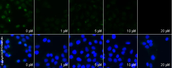
ab87359 staining cyclin A in DU145 cells treated with lovastatin (ab120614), by ICC/IF. Decrease in cyclin A expression correlates with increased concentration of lovastatin, as described in literature.The cells were incubated at 37°C for 24h in media containing different concentrations of ab120614 (lovastatin) in DMSO, fixed with 4% formaldehyde for 10 minutes at room temperature and blocked with PBS containing 10% goat serum, 0.3 M glycine, 1% BSA and 0.1% tween for 2h at room temperature. Staining of the treated cells with ab87359(5 µg/ml) was performed overnight at 4°C in PBS containing 1% BSA and 0.1% tween. A DyLight 488 goat anti-rabbit polyclonal antibody (ab96899) at 1/250 dilution was used as the secondary antibody. Nuclei were counterstained with DAPI and are shown in blue.