Anti-CYP27B1 antibody (ab95047) at 1/1000 dilution + Mouse kidney tissue lysate at 35 µgSecondaryPeroxidase-conjugated goat anti-rabbit IgG (H+L) at 1/5000 dilution

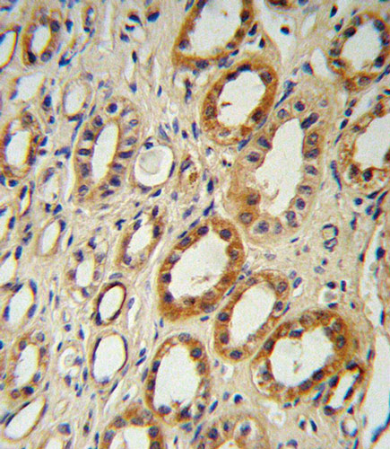
Immunohistochemistry (Formalin/PFA-fixed paraffin-embedded sections) analysis of human kidney tissue labelling CYP27B1 with ab95047. Tissue was fixed with formaldehyde and blocked with 3% BSA for 0.5 hour at 38°C; antigen retrieval was by heat mediation with a citrate buffer (pH6). Samples were incubated with primary antibody (1/25) for 1 hour at 37°C. A peroxidase-conjugated goat anti-rabbit polyclonal (ready to use) was used as the secondary antibody.

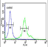
Flow Cytometry analysis of 293 cells labelling CYP27B1 with ab95047 (green) compared to a negative control (blue). The cells were fixed with paraformaldehyde and then permeabilized with 90% methanol for 10 min. The cells were then incubated in 3% BSA to block non-specific protein-protein interactions followed by the primary antibody (1µg/1x106 cells) for 60 min at 37ºC. The secondary antibody, a FITC conjugated goat anti-rabbit IgG, was used at 1/200 dilution for 40 min at room temperature. Acquisition of >10,000 events was performed.


