Anti-Cytochrome C Oxidase subunit VIc antibody [3G5F7G3]
| Name | Anti-Cytochrome C Oxidase subunit VIc antibody [3G5F7G3] |
|---|---|
| Supplier | Abcam |
| Catalog | ab110267 |
| Prices | $384.00 |
| Sizes | 100 µg |
| Host | Mouse |
| Clonality | Monoclonal |
| Isotype | IgG2b |
| Clone | 3G5F7G3 |
| Applications | ICC/IF ICC/IF FC WB |
| Species Reactivities | Rat, Bovine, Human |
| Antigen | Cytochrome C Oxidase subunit VIc from Bovine Heart |
| Description | Mouse Monoclonal |
| Gene | COX6C |
| Conjugate | Unconjugated |
| Supplier Page | Shop |
Product images
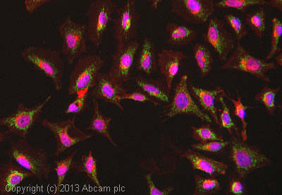 ICC/IF image of ab110267 stained HeLa cells. The cells were 4% formaldehyde fixed (10 min) and then incubated in 1%BSA / 10% normal goat serum / 0.3M glycine in 0.1% PBS-Tween for 1h to permeabilise the cells and block non-specific protein-protein interactions. The cells were then incubated with the antibody (ab110267, 5µg/ml) overnight at +4°C. The secondary antibody (green) was ab96879, DyLight® 488 goat anti-mouse IgG (H+L) used at a 1/250 dilution for 1h. Alexa Fluor® 594 WGA was used to label plasma membranes (red) at a 1/200 dilution for 1h. DAPI was used to stain the cell nuclei (blue) at a concentration of 1.43µM.
![All lanes : Anti-Cytochrome C Oxidase subunit VIc antibody [3G5F7G3] (ab110267) at 2 µg/mlLane 1 : Isolated mitochondria from Human heart at 50 µgLane 2 : Isolated mitochondria from Bovine heart at 1 µgLane 3 : Isolated mitochondria from Rat heart at 10 µg](http://www.bioprodhub.com/system/product_images/ab_products/2/sub_2/4756_Cytochrome-C-Oxidase-subunit-VIc-Primary-antibodies-ab110267-1.jpg) All lanes : Anti-Cytochrome C Oxidase subunit VIc antibody [3G5F7G3] (ab110267) at 2 µg/mlLane 1 : Isolated mitochondria from Human heart at 50 µgLane 2 : Isolated mitochondria from Bovine heart at 1 µgLane 3 : Isolated mitochondria from Rat heart at 10 µg
All lanes : Anti-Cytochrome C Oxidase subunit VIc antibody [3G5F7G3] (ab110267) at 2 µg/mlLane 1 : Isolated mitochondria from Human heart at 50 µgLane 2 : Isolated mitochondria from Bovine heart at 1 µgLane 3 : Isolated mitochondria from Rat heart at 10 µg
![Overlay histogram showing HepG2 cells stained with ab110267 (red line). The cells were fixed with 80% methanol (5 min) and then permeabilized with 0.1% PBS-Tween for 20 min. The cells were then incubated in 1x PBS / 10% normal goat serum / 0.3M glycine to block non-specific protein-protein interactions. The cells were then incubated with the antibody (ab110267, 1µg/1x106 cells) for 30 min at 22ºC. The secondary antibody used was DyLight® 488 goat anti-mouse IgG (H+L) (ab96879) at 1/500 dilution for 30 min at 22ºC. Isotype control antibody (black line) was mouse IgG2b [PLPV219] (ab91366, 2µg/1x106 cells) used under the same conditions. Acquisition of >5,000 events was performed. This antibody gave a positive signal in HepG2 cells fixed with 4% paraformaldehyde (10 min)/permeabilized with 0.1% PBS-Tween for 20 min used under the same conditions.](http://www.bioprodhub.com/system/product_images/ab_products/2/sub_2/4757_Cytochrome-C-Oxidase-subunit-VIc-Primary-antibodies-ab110267-2.jpg) Overlay histogram showing HepG2 cells stained with ab110267 (red line). The cells were fixed with 80% methanol (5 min) and then permeabilized with 0.1% PBS-Tween for 20 min. The cells were then incubated in 1x PBS / 10% normal goat serum / 0.3M glycine to block non-specific protein-protein interactions. The cells were then incubated with the antibody (ab110267, 1µg/1x106 cells) for 30 min at 22ºC. The secondary antibody used was DyLight® 488 goat anti-mouse IgG (H+L) (ab96879) at 1/500 dilution for 30 min at 22ºC. Isotype control antibody (black line) was mouse IgG2b [PLPV219] (ab91366, 2µg/1x106 cells) used under the same conditions. Acquisition of >5,000 events was performed. This antibody gave a positive signal in HepG2 cells fixed with 4% paraformaldehyde (10 min)/permeabilized with 0.1% PBS-Tween for 20 min used under the same conditions.
Overlay histogram showing HepG2 cells stained with ab110267 (red line). The cells were fixed with 80% methanol (5 min) and then permeabilized with 0.1% PBS-Tween for 20 min. The cells were then incubated in 1x PBS / 10% normal goat serum / 0.3M glycine to block non-specific protein-protein interactions. The cells were then incubated with the antibody (ab110267, 1µg/1x106 cells) for 30 min at 22ºC. The secondary antibody used was DyLight® 488 goat anti-mouse IgG (H+L) (ab96879) at 1/500 dilution for 30 min at 22ºC. Isotype control antibody (black line) was mouse IgG2b [PLPV219] (ab91366, 2µg/1x106 cells) used under the same conditions. Acquisition of >5,000 events was performed. This antibody gave a positive signal in HepG2 cells fixed with 4% paraformaldehyde (10 min)/permeabilized with 0.1% PBS-Tween for 20 min used under the same conditions.
Product References
Age-related decrease in expression of mitochondrial DNA encoded subunits of - Age-related decrease in expression of mitochondrial DNA encoded subunits of
Sohal RS, Toroser D, Bregere C, Mockett RJ, Orr WC. Mech Ageing Dev. 2008 Sep;129(9):558-61.
The A3243G tRNALeu(UUR) mutation induces mitochondrial dysfunction and variable - The A3243G tRNALeu(UUR) mutation induces mitochondrial dysfunction and variable
Janssen GM, Hensbergen PJ, van Bussel FJ, Balog CI, Maassen JA, Deelder AM, Raap AK. Hum Mol Genet. 2007 Oct 15;16(20):2472-81. Epub 2007 Jul 25.
The expression of polymerase gamma and mitochondrial transcription factor A and - The expression of polymerase gamma and mitochondrial transcription factor A and
Amaral A, Ramalho-Santos J, St John JC. Hum Reprod. 2007 Jun;22(6):1585-96. Epub 2007 Mar 5.
Immunodetecting members of the Bcl-2 family of proteins. - Immunodetecting members of the Bcl-2 family of proteins.
Lock RB, Murphy KM. Methods Mol Med. 2005;111:83-96.