Anti-Cytochrome P450 1A2 antibody
| Name | Anti-Cytochrome P450 1A2 antibody |
|---|---|
| Supplier | Abcam |
| Catalog | ab170204 |
| Prices | $376.00 |
| Sizes | 400 µl |
| Host | Rabbit |
| Clonality | Polyclonal |
| Isotype | IgG |
| Applications | ICC/IF ICC/IF IHC-P WB |
| Species Reactivities | Mouse, Human |
| Antigen | Synthetic peptide within Human Cytochrome P450 1A2 aa 255-282 (internal sequence) conjugated to Keyhole Limpet Haemocyanin (KLH) |
| Description | Rabbit Polyclonal |
| Gene | CYP1A2 |
| Conjugate | Unconjugated |
| Supplier Page | Shop |
Product images
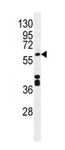 Anti-Cytochrome P450 1A2 antibody (ab170204) + Mouse liver tissue lysate at 35 µg
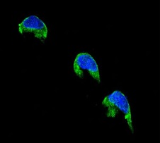 Immunocytochemistry/ Immunofluorescence analysis of 293 cells labelling Cytochrome P450 1A2 (green) with ab170204. An Alexa Fluor® 488-conjugated goat anti-rabbit IgG was used as the secondary antibody. Counterstained with DAPI (blue).
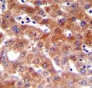 Immunohistochemistry (Formalin/PFA-fixed paraffin-embedded sections) analysis of human liver tissue labelling Cytochrome P450 1A2 with ab170204. A peroxidase-conjugated anti-rabbit IgG was used as the secondary antibody, followed by DAB staining.