Anti-Cytochrome P450 1A2 antibody [d15 (16VII F10F12)]
| Name | Anti-Cytochrome P450 1A2 antibody [d15 (16VII F10F12)] |
|---|---|
| Supplier | Abcam |
| Catalog | ab22717 |
| Prices | $392.00 |
| Sizes | 100 µg |
| Host | Mouse |
| Clonality | Monoclonal |
| Isotype | IgG1 |
| Clone | d15 (16VII F10F12) |
| Applications | FC IHC-P ICC/IF ICC/IF ELISA WB |
| Species Reactivities | Mouse, Rat, Human |
| Antigen | Full length protein (Rat) |
| Description | Mouse Monoclonal |
| Gene | CYP1A2 |
| Conjugate | Unconjugated |
| Supplier Page | Shop |
Product images
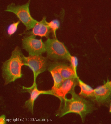 ICC/IF image of ab22717 stained MCF7 cells. The cells were 100% methanol fixed (5 min) and then incubated in 1%BSA / 10% normal goat serum / 0.3M glycine in 0.1% PBS-Tween for 1h to permeabilise the cells and block non-specific protein-protein interactions. The cells were then incubated with the antibody (ab22717, 1µg/ml) overnight at +4°C. The secondary antibody (green) was Alexa Fluor® 488 goat anti-mouse IgG (H+L) used at a 1/1000 dilution for 1h. Alexa Fluor® 594 WGA was used to label plasma membranes (red) at a 1/200 dilution for 1h. DAPI was used to stain the cell nuclei (blue) at a concentration of 1.43µM.
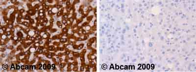 Ab22717 staining human normal liver. Staining is localised to the cytoplasm.Left panel: with primary antibody at 1 ug/ml. Right panel: isotype control.Sections were stained using an automated system DAKO Autostainer Plus , at room temperature. Sections were rehydrated and antigen retrieved with the Dako 3-in-1 antigen retrieval buffer citrate pH 6.0 in a DAKO PT Link. Slides were peroxidase blocked in 3% H2O2 in methanol for 10 minutes. They were then blocked with Dako Protein block for 10 minutes (containing casein 0.25% in PBS) then incubated with primary antibody for 20 minutes and detected with Dako Envision Flex amplification kit for 30 minutes. Colorimetric detection was completed with diaminobenzidine for 5 minutes. Slides were counterstained with Haematoxylin and coverslipped under DePeX. Please note that for manual staining we recommend to optimize the primary antibody concentration and incubation time (overnight incubation), and amplification may be required.
![All lanes : Anti-Cytochrome P450 1A2 antibody [d15 (16VII F10F12)] (ab22717) at 1 µg/mlLane 1 : Liver (Human) Tissue Lysate - adult normal tissue (ab29889)Lane 2 : Liver (Mouse) Tissue LysateLane 3 : Liver (Rat) Tissue LysateLysates/proteins at 10 µg per lane.SecondaryGoat Anti-Mouse IgG H&L (HRP) preadsorbed (ab97040) at 1/5000 dilutiondeveloped using the ECL techniquePerformed under reducing conditions.Observed band size : 58 kDa (why is the actual band size different from the predicted?)Additional bands at : 30 kDa,48 kDa. We are unsure as to the identity of these extra bands.Exposure time : 150 seconds](http://www.bioprodhub.com/system/product_images/ab_products/2/sub_2/4824_Cytochrome-P450-1A2-Primary-antibodies-ab22717-4.jpg) All lanes : Anti-Cytochrome P450 1A2 antibody [d15 (16VII F10F12)] (ab22717) at 1 µg/mlLane 1 : Liver (Human) Tissue Lysate - adult normal tissue (ab29889)Lane 2 : Liver (Mouse) Tissue LysateLane 3 : Liver (Rat) Tissue LysateLysates/proteins at 10 µg per lane.SecondaryGoat Anti-Mouse IgG H&L (HRP) preadsorbed (ab97040) at 1/5000 dilutiondeveloped using the ECL techniquePerformed under reducing conditions.Observed band size : 58 kDa (why is the actual band size different from the predicted?)Additional bands at : 30 kDa,48 kDa. We are unsure as to the identity of these extra bands.Exposure time : 150 seconds
All lanes : Anti-Cytochrome P450 1A2 antibody [d15 (16VII F10F12)] (ab22717) at 1 µg/mlLane 1 : Liver (Human) Tissue Lysate - adult normal tissue (ab29889)Lane 2 : Liver (Mouse) Tissue LysateLane 3 : Liver (Rat) Tissue LysateLysates/proteins at 10 µg per lane.SecondaryGoat Anti-Mouse IgG H&L (HRP) preadsorbed (ab97040) at 1/5000 dilutiondeveloped using the ECL techniquePerformed under reducing conditions.Observed band size : 58 kDa (why is the actual band size different from the predicted?)Additional bands at : 30 kDa,48 kDa. We are unsure as to the identity of these extra bands.Exposure time : 150 seconds
![All lanes : Anti-Cytochrome P450 1A2 antibody [d15 (16VII F10F12)] (ab22717) at 1/2500 dilutionLane 1 : Tissue lysate prepared from murine liver microsomesLane 2 : Tissue lysate prepared from murine liver microsomesLane 3 : Tissue lysate prepared from murine liver microsomesLane 4 : Tissue lysate prepared from murine liver microsomesLysates/proteins at 10 µg per lane.SecondaryGoat anti-mouse IgG(H+L)-HRP conjugate at 1/5000 dilutiondeveloped using the ECL techniqueExposure time : 1 secondImage courtesy of an anonymous Abreview.See Abreview](http://www.bioprodhub.com/system/product_images/ab_products/2/sub_2/4825_Cytochrome-P450-1A2-Primary-antibodies-ab22717-13.jpg) All lanes : Anti-Cytochrome P450 1A2 antibody [d15 (16VII F10F12)] (ab22717) at 1/2500 dilutionLane 1 : Tissue lysate prepared from murine liver microsomesLane 2 : Tissue lysate prepared from murine liver microsomesLane 3 : Tissue lysate prepared from murine liver microsomesLane 4 : Tissue lysate prepared from murine liver microsomesLysates/proteins at 10 µg per lane.SecondaryGoat anti-mouse IgG(H+L)-HRP conjugate at 1/5000 dilutiondeveloped using the ECL techniqueExposure time : 1 secondImage courtesy of an anonymous Abreview.See Abreview
All lanes : Anti-Cytochrome P450 1A2 antibody [d15 (16VII F10F12)] (ab22717) at 1/2500 dilutionLane 1 : Tissue lysate prepared from murine liver microsomesLane 2 : Tissue lysate prepared from murine liver microsomesLane 3 : Tissue lysate prepared from murine liver microsomesLane 4 : Tissue lysate prepared from murine liver microsomesLysates/proteins at 10 µg per lane.SecondaryGoat anti-mouse IgG(H+L)-HRP conjugate at 1/5000 dilutiondeveloped using the ECL techniqueExposure time : 1 secondImage courtesy of an anonymous Abreview.See Abreview
![Overlay histogram showing MCF7 cells stained with ab22717 (red line). The cells were fixed with 80% methanol (5 min) and then permeabilized with 0.1% PBS-Tween for 20 min. The cells were then incubated in 1x PBS / 10% normal goat serum / 0.3M glycine to block non-specific protein-protein interactions followed by the antibody (ab22717, 1µg/1x106 cells) for 30 min at 22ºC. The secondary antibody used was DyLight® 488 goat anti-mouse IgG (H+L) (ab96879) at 1/500 dilution for 30 min at 22ºC. Isotype control antibody (black line) was mouse IgG1 [ICIGG1] (ab91353, 2µg/1x106 cells) used under the same conditions. Acquisition of >5,000 events was performed.](http://www.bioprodhub.com/system/product_images/ab_products/2/sub_2/4826_Cytochrome-P450-1A2-Primary-antibodies-ab22717-14.jpg) Overlay histogram showing MCF7 cells stained with ab22717 (red line). The cells were fixed with 80% methanol (5 min) and then permeabilized with 0.1% PBS-Tween for 20 min. The cells were then incubated in 1x PBS / 10% normal goat serum / 0.3M glycine to block non-specific protein-protein interactions followed by the antibody (ab22717, 1µg/1x106 cells) for 30 min at 22ºC. The secondary antibody used was DyLight® 488 goat anti-mouse IgG (H+L) (ab96879) at 1/500 dilution for 30 min at 22ºC. Isotype control antibody (black line) was mouse IgG1 [ICIGG1] (ab91353, 2µg/1x106 cells) used under the same conditions. Acquisition of >5,000 events was performed.
Overlay histogram showing MCF7 cells stained with ab22717 (red line). The cells were fixed with 80% methanol (5 min) and then permeabilized with 0.1% PBS-Tween for 20 min. The cells were then incubated in 1x PBS / 10% normal goat serum / 0.3M glycine to block non-specific protein-protein interactions followed by the antibody (ab22717, 1µg/1x106 cells) for 30 min at 22ºC. The secondary antibody used was DyLight® 488 goat anti-mouse IgG (H+L) (ab96879) at 1/500 dilution for 30 min at 22ºC. Isotype control antibody (black line) was mouse IgG1 [ICIGG1] (ab91353, 2µg/1x106 cells) used under the same conditions. Acquisition of >5,000 events was performed.
Product References
Quantitative proteomic and functional analysis of liver mitochondria from high - Quantitative proteomic and functional analysis of liver mitochondria from high
Guo Y, Darshi M, Ma Y, Perkins GA, Shen Z, Haushalter KJ, Saito R, Chen A, Lee YS, Patel HH, Briggs SP, Ellisman MH, Olefsky JM, Taylor SS. Mol Cell Proteomics. 2013 Dec;12(12):3744-58.
Susceptibility to acetaminophen (APAP) toxicity unexpectedly is decreased during - Susceptibility to acetaminophen (APAP) toxicity unexpectedly is decreased during
Getachew Y, James L, Lee WM, Thiele DL, Miller BC. Biochem Pharmacol. 2010 May 1;79(9):1363-71.
Multiple-approaches to the identification and quantification of cytochromes P450 - Multiple-approaches to the identification and quantification of cytochromes P450
Seibert C, Davidson BR, Fuller BJ, Patterson LH, Griffiths WJ, Wang Y. J Proteome Res. 2009 Apr;8(4):1672-81.
Comparative cytochrome P450 proteomics in the livers of immunodeficient mice - Comparative cytochrome P450 proteomics in the livers of immunodeficient mice
Lane CS, Wang Y, Betts R, Griffiths WJ, Patterson LH. Mol Cell Proteomics. 2007 Jun;6(6):953-62. Epub 2007 Feb 11.