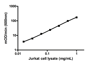
Example standard curve. A dilution series of recombinant CTIP2 in the working range of the assay.

Example of serially diluted endogenous CTIP2 expression in Jurkat cells.

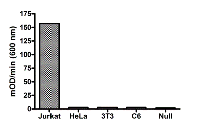
Demonstration of the specificity of this ELISA. Endogenous expression of CTIP2 in Jurkat cells is higher than other non-lymphocyte cells such as Hela (human carcinoma) and NIH3T3 (mouse fibroblast), and C6 (rat glioma).

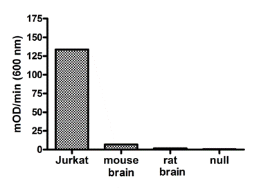
Demonstration of the specificity of this ELISA. CTIP2 expression is detectable in mouse brain lysate but not in rat brain lysate.

Demonstration of the specificity of the detector antibody used in this kit. (A) Western blot using the CTIP2 detector antibody at 1 µg/mL: Jurkat cell lysate and HeLa cell lysate, each were loaded at 16 µg/lane. (B) Analysis of CTIP2 staining in Jurkat cells by ICC: Left panel DAPI (red for contrast) with no primary antibody. Right panel CTIP2 detector primary antibody at 1 µg/mL (green), both samples contain 488 labeled secondary antibody. (C) Analysis of CTIP2 staining in Jurkat cells by Flow: Black unstained, red no primary and blue CTIP2 detector antibody at 1 µg/mL, red and blue include 488 dye labeled secondary.


